
Separation device and method thereof

A separation device and sample collection technology, applied in the pretreatment field of cytological samples, can solve the problems of pathological diagnosis and detection difficulties, failure to maintain cell shape, cell necrosis and disintegration, etc., to avoid pollution and reproduction, clear cells, reduce The effect of the false negative rate

Pending Publication Date: 2022-02-01
昂佰生物医学科技(苏州)有限公司
View PDF0 Cites 0 Cited by
  • Summary
  • Abstract
  • Description
  • Claims
  • Application Information

AI Technical Summary

Problems solved by technology

Since there are a lot of protein and other components in the pleural fluid, it is in a non-sterile state after collection, and it is very easy for bacteria to grow and contaminate the sample.
If the isolated cell samples are not fixed in time, the cells will be necrotic and disintegrated rapidly, the cell shape cannot be maintained, and macromolecules such as DNA and protein will also degrade and disappear, which brings great difficulties to pathological diagnosis and detection, and even gives false negative results. It is a big problem that has plagued clinical experimental medicine for a long time

Method used


Image

  • Separation device and method thereof
  • Separation device and method thereof
  • Separation device and method thereof

Examples


Embodiment 1

[0064] Example 1 Separation of Cells from Pleural and Ascites Samples

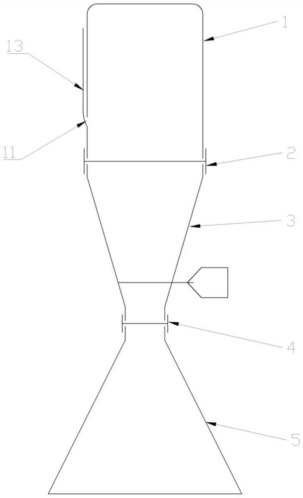
[0065] The separation device of the present invention is used to separate cells in a pleural and ascites sample, and the separation device includes a sample collector, a filter, a connector, a catcher and a filtrate collector.

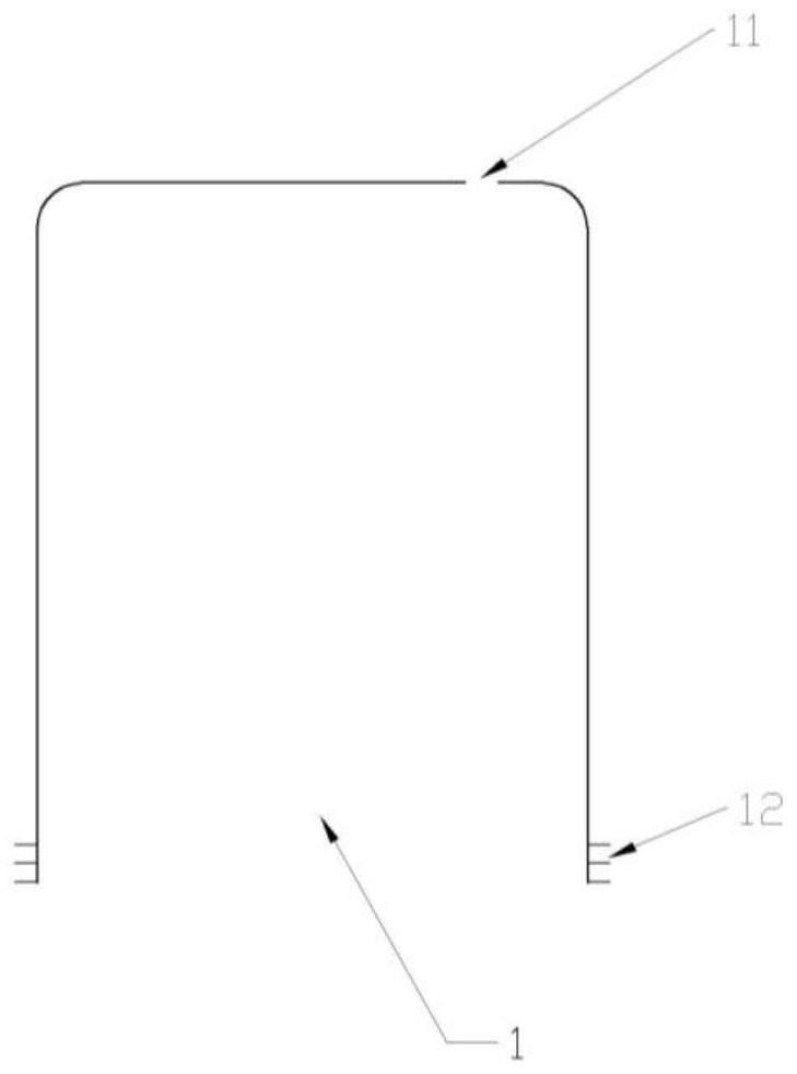
[0066] The sample collector is a plastic bottle with a screw mouth. There is an air inlet hole with a diameter of 5mm at the bottom of the bottle. The hole is covered and sealed with a plastic film. The air inlet hole can be pierced with a sharp instrument. The volume of the sample collector is 1000ml, and the diameter of the screw mouth is 100mm.

[0067] Filters shaped like Figure 4 , there is a spiral on the inner surface, which matches the bottle mouth of the sample collector and can be screwed tightly to seal. The filter is in the middle of the filter. can be outside. The diameter of the screw opening on one side of the filter is corresponding to the screw opening of the s...

Embodiment 2

[0075] Example 2 Separation of cells from stool samples

[0076] The separation device of the present invention is used to separate cells in a stool sample, and the separation device includes a sample collector, a filter, a connector, a catcher and a filtrate collector.

[0077] The sample collector is an open funnel-shaped container, and the lower opening of the funnel is provided with a buckle, which is connected with the filter.

[0078] Filters shaped like Figure 4 , there is a buckle on the inner surface, which is buckled and sealed with the lower mouth of the sample collection container. The filter is in the middle of the filter, and there is also a buckle on the opposite side of the filter and the sample collector. The filter can be divided into different models according to the filter pore size, preferably divided into three types: coarse, medium and fine. The pore size of the coarse pore model is 3mm, the pore size of the medium hole is 1.5mm, and the pore size of t...


PUM

PropertyMeasurementUnit
volumeaaaaaaaaaa
diameteraaaaaaaaaa
lengthaaaaaaaaaa

Abstract

The invention discloses a separation device, which comprises a sample collector, a filter, a connector, a capturer and a filtrate collector, the sample collector and the filtrate collector are both containers, the filter comprises a filter screen, the capturer comprises a capture chip with the aperture of 0.01-1000 [mu] m, the connector is a barrel-shaped object, and the sample collector, the filter, the connector, the capturer and the filtrate collector are connected in sequence. According to the separating device disclosed by the invention, before samples such as clinical hydrothorax and ascites, urine and the like are transferred to a laboratory for detection, particulate matters to be detected in the samples can be separated and fixed at the same time at a sampling site, the intact forms of cells are kept, the cells are prevented from disintegration and degradation of biomacromolecules in the cells are prevented, and microbial pollution and reproduction are avoided; the false negative rate caused by the storage and transportation process of the sample is effectively reduced, and the detection rate of tumor cells is greatly improved.

Description

technical field [0001] The invention relates to the technical field of pretreatment of cytological samples, in particular to a device and method for separating exfoliated cells or microorganisms in samples such as body fluids and excrement in medical diagnosis and treatment. Background technique [0002] Clinically, early diagnosis and early treatment of tumors are very important. Especially for lung cancer, the first clinical symptom in many cases is pleural effusion. For colon cancer and urinary tract tumors, colonoscopy and cystoscopy are routine diagnostic methods, but the operation is complicated, the patients are painful, and they are not suitable for screening. As a non-invasive, low-cost and convenient stool and urine examination, it has become the preferred method. However, these samples were not processed in time before they were sent to the laboratory, and when they were checked by the laboratory, especially for samples far away from medical laboratories, they ne...

Claims


Application Information

Patent Timeline
Patent Type & Authority Applications(China)
IPC IPC(8): C12M1/12C12M1/26
CPCC12M33/14
Inventor 张秀梅谢玲饶娟娟
Owner 昂佰生物医学科技(苏州)有限公司
Learn More