
Fixed observation device for animal spinal nerve living body imaging and use method thereof

An observation device and in vivo imaging technology, applied in the field of medical devices, can solve the problems of difficult to achieve long-term observation and research, difficult to observe the mutation of the same axis, and anatomical complexity, so as to improve the accuracy of observation, avoid incision infection, Wide range of effects

Pending Publication Date: 2021-09-21
SHENZHEN PEOPLES HOSPITAL
View PDF11 Cites 2 Cited by
  • Summary
  • Abstract
  • Description
  • Claims
  • Application Information

AI Technical Summary

Benefits of technology

This patented technology allows users to see their own brains through an opaque chamber with both eyes open at once instead of repeatedly cutting them down when observing it over longer periods or even after being taken outdoors due to its ability to prevent bacteria growth around joints without requiring invasive surgeries that may cause pain or damage. Additionally, this device provides stable motion control while still allowing us to monitor changes made about how much tissue needs repair within our bodies. Overall, these technical features make studying Brain Nerves (BN) more accessible and easier than previous methods such as histopathology techniques.

Problems solved by technology

This patented method involves traditional methods that involve exposure during surgeries or injections into tissue around the spine. These techniques can be challenged by limitations such as lack of space between neurons and difficulties with fixing them properly over longer periods of observation (such as prolonged visualization).

Method used


Image

  • Fixed observation device for animal spinal nerve living body imaging and use method thereof
  • Fixed observation device for animal spinal nerve living body imaging and use method thereof
  • Fixed observation device for animal spinal nerve living body imaging and use method thereof

Examples


Embodiment Construction

[0031] The present invention will be described in further detail below in conjunction with the accompanying drawings.

[0032] This specific embodiment is only an explanation of the present invention, and it is not a limitation of the present invention. Those skilled in the art can make modifications to this embodiment as required after reading this specification, but as long as they are within the rights of the present invention All claims are protected by patent law.

[0033] The fixed observation device for live imaging of animal spinal nerves of the present invention realizes long-term (multiple) living dynamic observation and tracking of spinal nerves, and opens up new horizons for the research on peripheral nerve regeneration and neuropathic pain mechanisms.

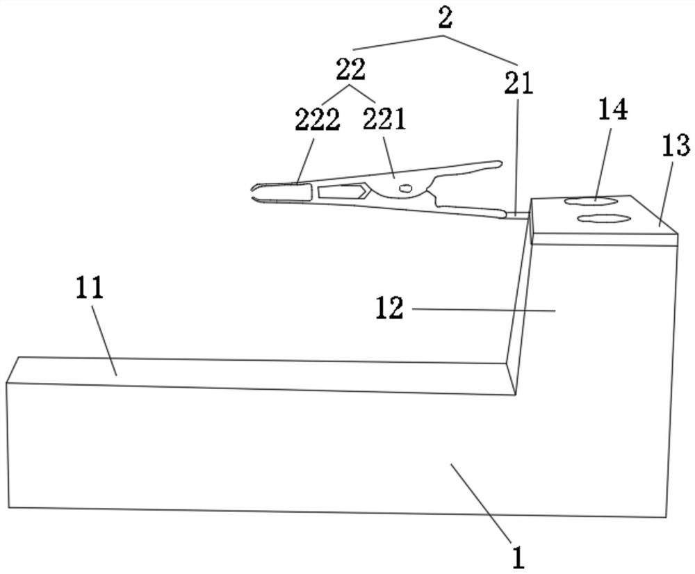
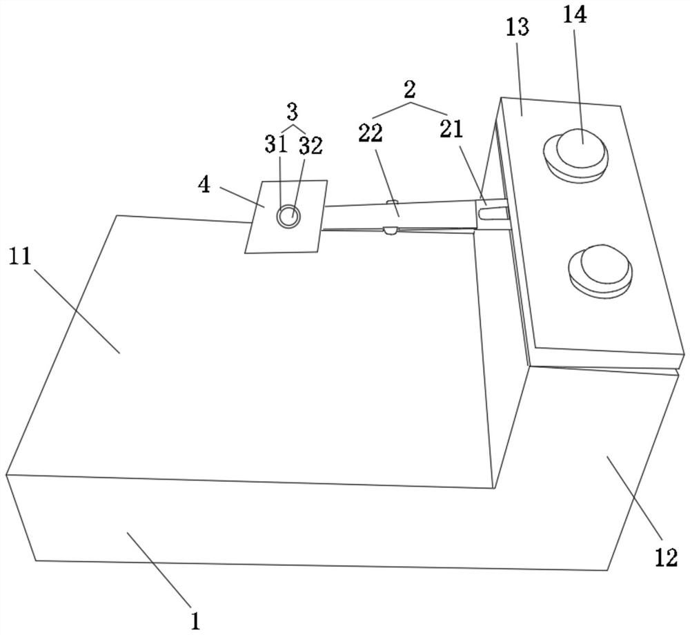
[0034] This embodiment relates to a fixed observation device for live imaging of animal spinal nerves. Such as figure 1 and figure 2 As shown, it includes: a base 1 , a fixing device 2 and an observation device...


PUM


Abstract

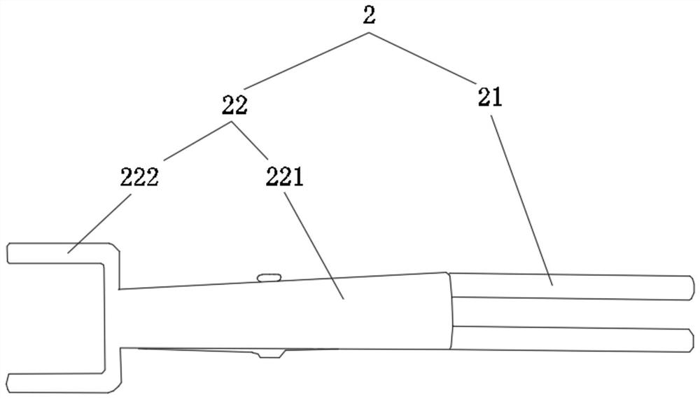
The invention discloses a fixed observation device for animal spinal nerve living body imaging and a use method thereof, and relates to the technical field of medical instruments. The device comprises a base, a fixing device and an observation device, the base comprises a base for placing an animal and a supporting piece fixedly arranged on the base; the fixing device comprises a fixing rod and a clamping piece, one end of the fixing rod is detachably fixed to the upper end of the supporting piece, and the clamping piece is arranged at the other end of the fixing rod and used for fixing the animal spine centrum; the observation device comprises an observation frame fixed on the clamping piece and an observation window arranged on the observation frame. By the adoption of the technical scheme, the device has the advantages of being good in fixing effect, capable of achieving long-time observation and capable of avoiding multiple operations.

Description


Claims


Application Information

Patent Timeline
Owner SHENZHEN PEOPLES HOSPITAL
Learn More