Application of Bmal1 gene in preparation of product for detecting and treating cartilage hypoplasia disease
A kind of achondroplasia and genetic technology, applied in the field of biomedicine, can solve the problems of lack of detection and treatment, and unsatisfactory curative effect
- Summary
- Abstract
- Description
- Claims
- Application Information
AI Technical Summary
Problems solved by technology
Method used
Image
Examples
Embodiment 1
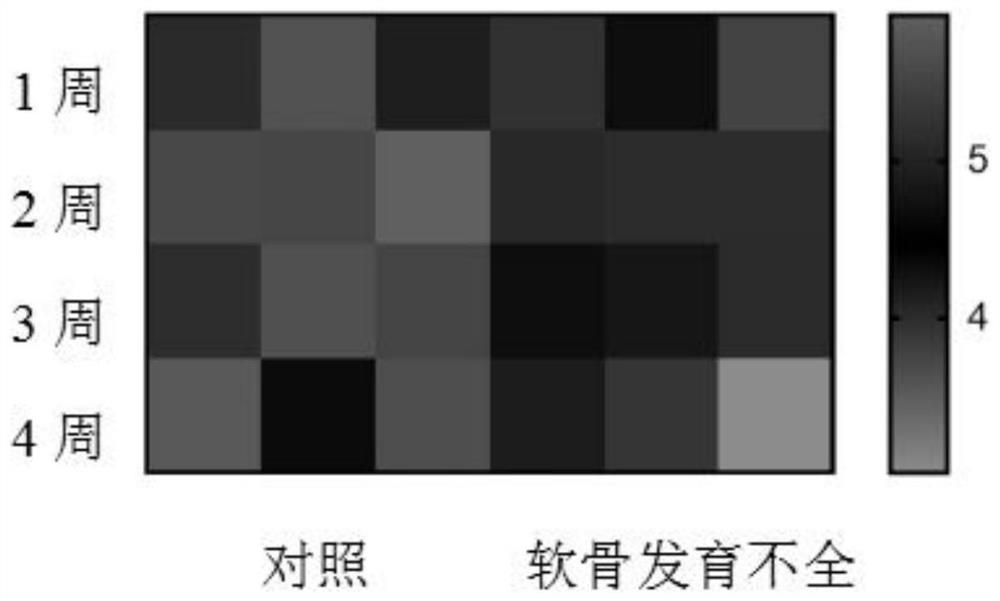
[0023] We downloaded the expression profile chips (GSE145821) of achondroplasia mice and their control mice through the GEO database, extracted and further analyzed the expression of the biological clock gene Bmal1, and found that in the femur tissues of 1-4 week achondroplasia mice, Compared with the control group, the expression of Bmal1 decreased, suggesting that there is a correlation between the decrease of Bmal1 gene and achondroplasia (see figure 1 ).
Embodiment 2
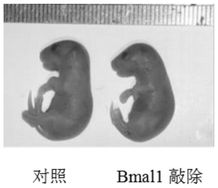
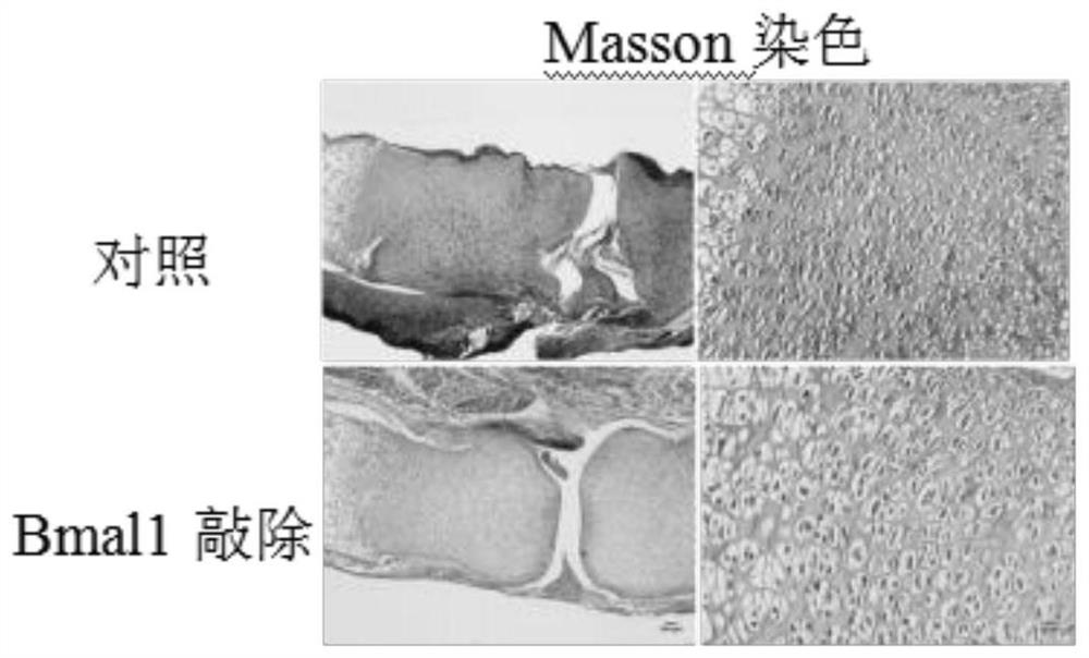
[0025] We bred Bmal1-knockout mice, and compared them with littermate wild-type mice, we found that Bmal1-knockout mice were shorter than control mice at embryonic day 18.5 (see figure 2 ), the results of Masson staining showed that the amount of collagen fibers in the femoral metaphysis cartilage tissue of Bmal1 knockout mice decreased, and the results of transmission electron microscopy showed that the endoplasmic reticulum in the femoral metaphysis chondrocytes of Bmal1 knockout mice was significantly expanded swelling, suggesting that the formation of cartilage matrix is regulated by Bmal1 (see image 3 , Figure 4 ).
Embodiment 3
[0027] We further extracted primary chondrocytes, knocked out Bmal1 in chondrocytes by infecting viruses, and detected the co-localization of chondrocyte type II collagen and endoplasmic reticulum by immunofluorescence experiments. The specific steps are as follows:
[0028] (1) Primary chondrocyte culture
[0029] 1) SD male rats, 60-80g, 2-3 weeks old, were killed by neck dislocation, and soaked in alcohol for 15 minutes;
[0030] 2) Take out the rat femur, carefully separate the femoral metaphyseal cartilage, and soak it with PBS;
[0031] 3) Transfer the cartilage tissue to an EP tube and cut it into pieces, add 1.5 mL of 0.25% trypsin, mix well, and place it in a 37°C water bath for 30-45 minutes to digest;
[0032] 4) Centrifuge the digested cartilage tissue at 1000 rpm for 5 minutes, discard the supernatant, add 8 mL of 0.25% type II collagenase prepared with basal medium, resuspend in a 15 mL centrifuge tube, and digest in an incubator at 37°C for 8-12 hours;
[0033...
PUM
 Abstract
Description
Claims
Application Information
 - R&D Engineer
- R&D Manager
- IP Professional
- Industry Leading Data Capabilities
- Powerful AI technology
- Patent DNA Extraction
Browse by: Latest US Patents, China's latest patents, Technical Efficacy Thesaurus, Application Domain, Technology Topic, Popular Technical Reports.
© 2024 PatSnap. All rights reserved.Legal|Privacy policy|Modern Slavery Act Transparency Statement|Sitemap|About US| Contact US: help@patsnap.com


