
3D printed bone defect repair scaffold loaded with endothelial extracellular matrix and preparation method of 3D printed bone defect repair scaffold

A technology of endothelial cells and 3D printing, which is applied in prosthetics, tissue regeneration, additive processing, etc., can solve problems such as short half-life, and achieve the effects of simple operation, increased number of branches, and high biological activity

Active Publication Date: 2021-06-18
STOMATOLOGY AFFILIATED STOMATOLOGY HOSPITAL OF GUANGZHOU MEDICAL UNIV
View PDF2 Cites 6 Cited by
  • Summary
  • Abstract
  • Description
  • Claims
  • Application Information

AI Technical Summary

Problems solved by technology

Adding growth factors such as bone matrix protein-2 (one matrix protein-2, BMP-2) to the composite scaffold can promote the osteogenic differentiation of stem cells, but due to its short half-life in vivo, in order to maintain an effective dose for a long time, it needs to be in the scaffold Added in a large amount, exceeding the safe standard dose of 1.5mg / ml, thus causing a series of adverse reactions, such as inflammation, heterotopic bone and tumor formation

Method used


Image

  • 3D printed bone defect repair scaffold loaded with endothelial extracellular matrix and preparation method of 3D printed bone defect repair scaffold
  • 3D printed bone defect repair scaffold loaded with endothelial extracellular matrix and preparation method of 3D printed bone defect repair scaffold
  • 3D printed bone defect repair scaffold loaded with endothelial extracellular matrix and preparation method of 3D printed bone defect repair scaffold

Examples


Embodiment 1

[0042] Preparation of a 3D printed bone defect repair scaffold loaded with endothelial extracellular matrix:

[0043] S1. Gelatin, sodium alginate and 58S bioglass were dissolved in distilled water to obtain a solution, wherein the mass / volume concentration of each component in the solution was gelatin 15%, sodium alginate 6%, and 58S bioglass 10.5%.

[0044] S2. The solution is stirred evenly by means of magnetic stirring and mechanical stirring to obtain 3D printing slurry, inject the 3D printing slurry into the 3D printing barrel, and start 3D printing after defoaming and homogenization. 3D printing uses a needle with an aperture of 0.41mm. Under the conditions of 0.38Mpa air pressure and 28°C, print at a printing speed of 10mm / s.

[0045] S3. The semi-finished stent is obtained after printing. The semi-finished stent is first physically cross-linked with a calcium chloride solution, and then chemically cross-linked by soaking in a glutaraldehyde solution; finally cleaned a...

Embodiment 2

[0050] Preparation of a 3D printed bone defect repair scaffold loaded with endothelial extracellular matrix:

[0051] S1. Gelatin, sodium alginate and 58S bioglass were dissolved in distilled water to obtain a solution, wherein the mass / volume concentration of each component in the solution was gelatin 15%, sodium alginate 6%, and 58S bioglass 10.5%.

[0052] S2. The solution is stirred evenly by means of magnetic stirring and mechanical stirring to obtain 3D printing slurry, inject the 3D printing slurry into the 3D printing barrel, and start 3D printing after defoaming and homogenization. 3D printing uses a needle with an aperture of 0.43mm. Under the conditions of 0.40Mpa air pressure and 27°C, print at a printing speed of 10mm / s.

[0053] S3. The semi-finished stent is obtained after printing. The semi-finished stent is first physically cross-linked with a calcium chloride solution, and then chemically cross-linked by soaking in a glutaraldehyde solution; finally cleaned a...

Embodiment 3

[0058] Preparation of a 3D printed bone defect repair scaffold loaded with endothelial extracellular matrix:

[0059] S1. Gelatin, sodium alginate and 58S bioglass were dissolved in distilled water to obtain a solution, wherein the mass / volume concentration of each component in the solution was gelatin 15%, sodium alginate 6%, and 58S bioglass 10.5%.

[0060] S2. Stir the solution evenly by magnetic stirring and mechanical stirring to obtain a 3D printing slurry, and prepare a bracket by 3D printing; in the 3D printing operation, inject the 3D printing slurry into the 3D printing barrel, start printing after defoaming and homogenization, Using a needle with an aperture of 0.40mm, print at a printing speed of 10mm / s under the conditions of 0.35Mpa air pressure and 29°C.

[0061] S3. The semi-finished stent is obtained after printing. The semi-finished stent is first physically cross-linked with a calcium chloride solution, and then chemically cross-linked by soaking in a glutar...


PUM

PropertyMeasurementUnit
pore sizeaaaaaaaaaa
pore sizeaaaaaaaaaa
pore sizeaaaaaaaaaa

Abstract

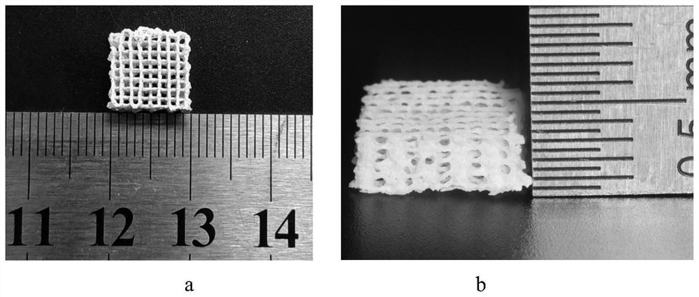
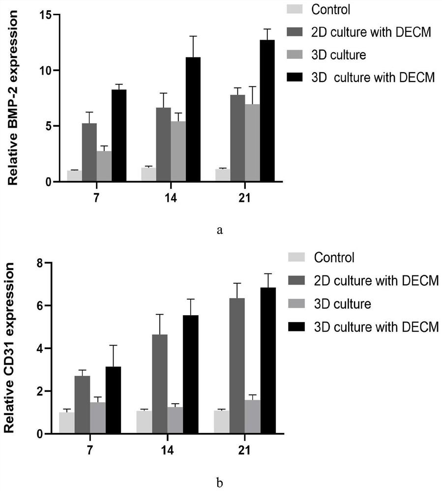
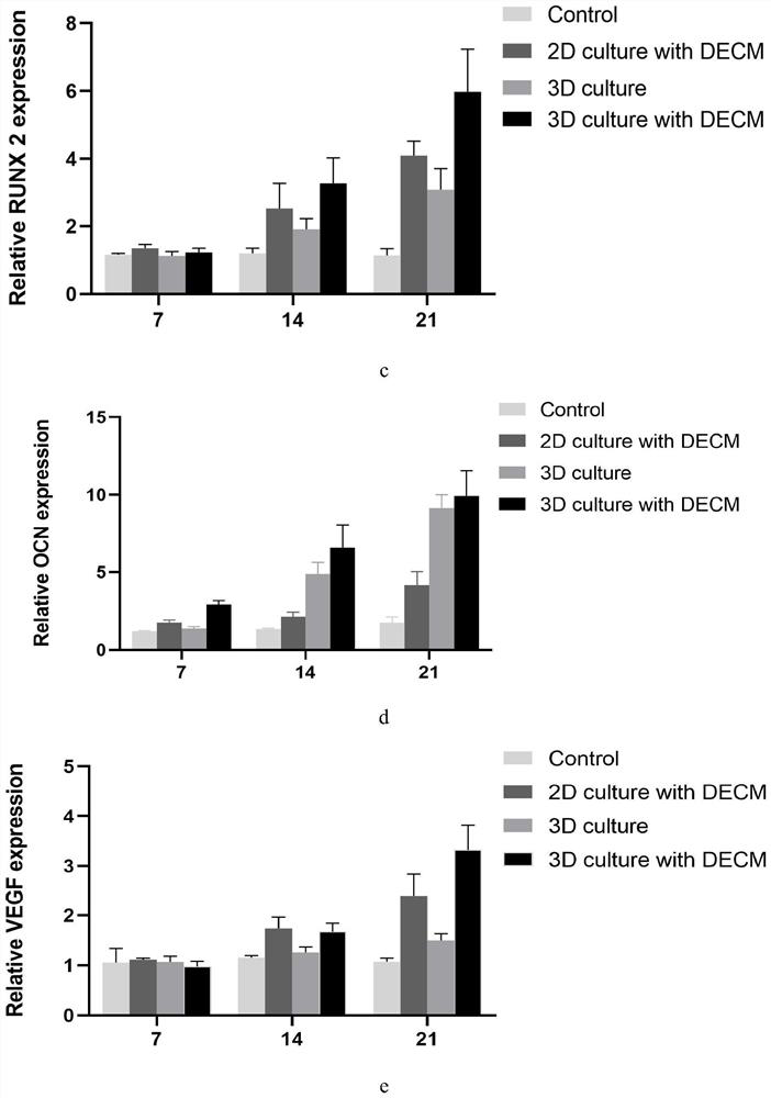
The invention discloses a 3D printed bone defect repair scaffold loaded with an endothelial extracellular matrix. The scaffold is prepared from raw materials including gelatin, sodium alginate and 58S bioglass, the pore diameter of the scaffold is 0.5-0.7 mm, and the porosity of the scaffold is 60-75%. A preparation process of the scaffold loaded with the endothelial extracellular matrix comprises the following steps: S1, performing disinfection treatment on a 3D printed scaffold; S2, inoculating the disinfected scaffold with RAOEC cells, changing the liquid every 3 days, and co-culturing the cells for 14 days; and S3, taking out the scaffold, and carrying out decellularization treatment and freeze-drying to obtain the scaffold. The scaffold is composed of the gelatin, the sodium alginate and the 58S bioglass, and is prepared by adopting a 3D printing technology, the operation is simple, and the structure and architecture of the scaffold are controllable; It is found that the extracellular matrix loaded withendothelial cells can promote osteogenesis and vascularization differentiation, the scaffold loaded with the extracellular matrix can promote formation of bone tissue and vascular tissue at the defect part, and the bone defect repair efficiency is obviously improved.

Description

technical field [0001] The invention belongs to the technical field of bone tissue engineering repair and reconstruction, and mainly relates to a 3D printed bone defect repair bracket loaded with endothelial extracellular matrix and a preparation method thereof. Background technique [0002] With the increase of bone tissue damage caused by aging, joint degeneration, traffic accidents and other trauma, more and more attention has been paid to the repair of bone defects. In clinical practice, bone transplantation methods such as autologous bone transplantation, allogeneic bone transplantation and artificial bone transplantation are usually used. Autologous bone transplantation is the "gold standard" for defect repair, but the source of autologous bone is limited, and the supply is often in short supply. Allogeneic bone grafts have the risk of infection and disease, while artificial bone grafts lack osteoinductive activity, and the osteogenesis efficiency is poor, making it dif...

Claims


Application Information

Patent Timeline
Patent Type & Authority Applications(China)
IPC IPC(8): A61L27/10A61L27/22A61L27/20A61L27/50A61L27/36A61L27/54B33Y70/00B33Y80/00B33Y10/00
CPCA61L27/10A61L27/222A61L27/20A61L27/50A61L27/3633A61L27/365A61L27/54B33Y70/00B33Y80/00B33Y10/00A61L2430/02A61L2300/10A61L2300/412A61L2430/40C08L5/04
Inventor 江千舟涂欣冉郭吕华张阳郭黎洋谭国忠陈荣丰
Owner STOMATOLOGY AFFILIATED STOMATOLOGY HOSPITAL OF GUANGZHOU MEDICAL UNIV
Learn More