Novel antigen epitope based on CD271 and application thereof
An antigen epitope, a new type of technology, applied in the biological field, can solve the problems of binding influence, shedding, undetectable, etc., and achieve the effect of stable binding, strong affinity and low homology
- Summary
- Abstract
- Description
- Claims
- Application Information
AI Technical Summary
Problems solved by technology
Method used
Image
Examples
Embodiment 1
[0062] [Example 1] Design, expression and purification of human CD271 antigen epitope peptide plasmid
[0063] 1. Selection of human CD271 epitope peptide
[0064] According to the full-length amino acid sequence of CD271 protein retrieved by NCBI, DNAStar software, IEDB website, etc. were used to analyze the antigenic determinant, and finally the epitope peptide was determined to be located in the SEQ ID NO.1 of the outer Stalk region of CD271 (named CD271T). Among them, a series of epitope peptides are designed to meet:
[0065] (1) located in the polypeptide chain SEQ ID NO.1; or,
[0066] (2) An amino acid sequence at least 80% identical to the sequence of SEQ ID NO. 1 in (1).
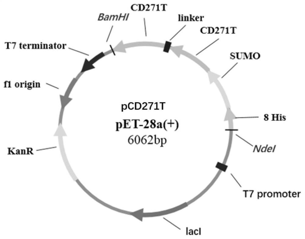
[0067] 2. Design of CD271T expression plasmid
[0068] According to the NCBI retrieval of the complete gene sequence of CD271 protein and the amino acid sequence of CD271T, the prokaryotic expression codon was optimized by Suzhou Synbio Co., Ltd., and cloned into the pET-28a(+) vector together w...
Embodiment 2
[0089] [Example 2] Proteolytic cleavage reaction and purification of human CD271T antigen peptide
[0090] 1. Protease digestion reaction and SDS-PAGE identification of human CD271T antigen peptide
[0091] (1) enzyme digestion system
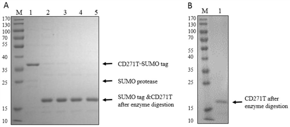
[0092] The enzyme digestion system is shown in Table 1, reacted in 30°C water bath for 1h, 2h, 4h, 6h, and carried out SDS-PAGE identification.
[0093] Table 1
[0094]
[0095]
[0096] (2) SDS-PAGE identification
[0097] Take 20 μL samples before enzyme digestion and 1h, 2h, 4h, and 6h after enzyme digestion, add 5×reduced SDS-PAGE loading buffer, heat at 95°C for 10 minutes, and centrifuge at 12,000 rpm for 5 minutes. Take 10 μL of each prepared sample for SDS-PAGE gel electrophoresis detection. For specific results, see image 3 Figure A in .
[0098] 2. Human CD271 immune antigen purification
[0099] Expand the enzyme digestion system according to the above reaction system, react in a water bath at 20°C for 2 hours, filter t...
Embodiment 3
[0100] [Example 3] Using the antigenic peptide in Example 1 to screen antibodies
[0101] 1. Antigen Preparation
[0102] 1) Antigen preparation for priming: Take a 1 mL sterile EP tube, dilute 100 μg of the immunogen with PBS to 100 μL and put it in the EP tube; shake well Freund’s complete adjuvant to mix the precipitated bifidobacteria evenly; take 100 μL Freund’s complete adjuvant Add the complete adjuvant to a 5mL sterile EP tube, then add the diluted immunogen, so that the volume ratio of the antigen to the adjuvant is 1:1, put the centrifuge tube in the ice box; prepare the ultrasonic breaker, Place the centrifuge tube in an ice box for sonication. Ultrasound conditions are total power 200W, total time 10min, working time 5 seconds, interval time 6 seconds. Observe the emulsification of the solution in real time; inhale the emulsified antigen with a 1 ml disposable syringe, and place it in an ice box for later use.
[0103] 2) Antigen preparation for the second and t...
PUM
 Abstract
Description
Claims
Application Information
 - R&D
- Intellectual Property
- Life Sciences
- Materials
- Tech Scout
- Unparalleled Data Quality
- Higher Quality Content
- 60% Fewer Hallucinations
Browse by: Latest US Patents, China's latest patents, Technical Efficacy Thesaurus, Application Domain, Technology Topic, Popular Technical Reports.
© 2025 PatSnap. All rights reserved.Legal|Privacy policy|Modern Slavery Act Transparency Statement|Sitemap|About US| Contact US: help@patsnap.com


