Rapid screening of tissue sample of rectal cancer aptamer and application of rectal cancer aptamer to preparation of detection preparation
A nucleic acid aptamer and rectal cancer technology, applied in DNA preparation, measurement devices, recombinant DNA technology, etc., to achieve low immunogenicity, good stability, and strong tissue permeability
- Summary
- Abstract
- Description
- Claims
- Application Information
AI Technical Summary
Problems solved by technology
Method used
Image
Examples
Embodiment 1
[0023]Example 1: Clinical rectal cancer tissue nucleic acid spiral screening
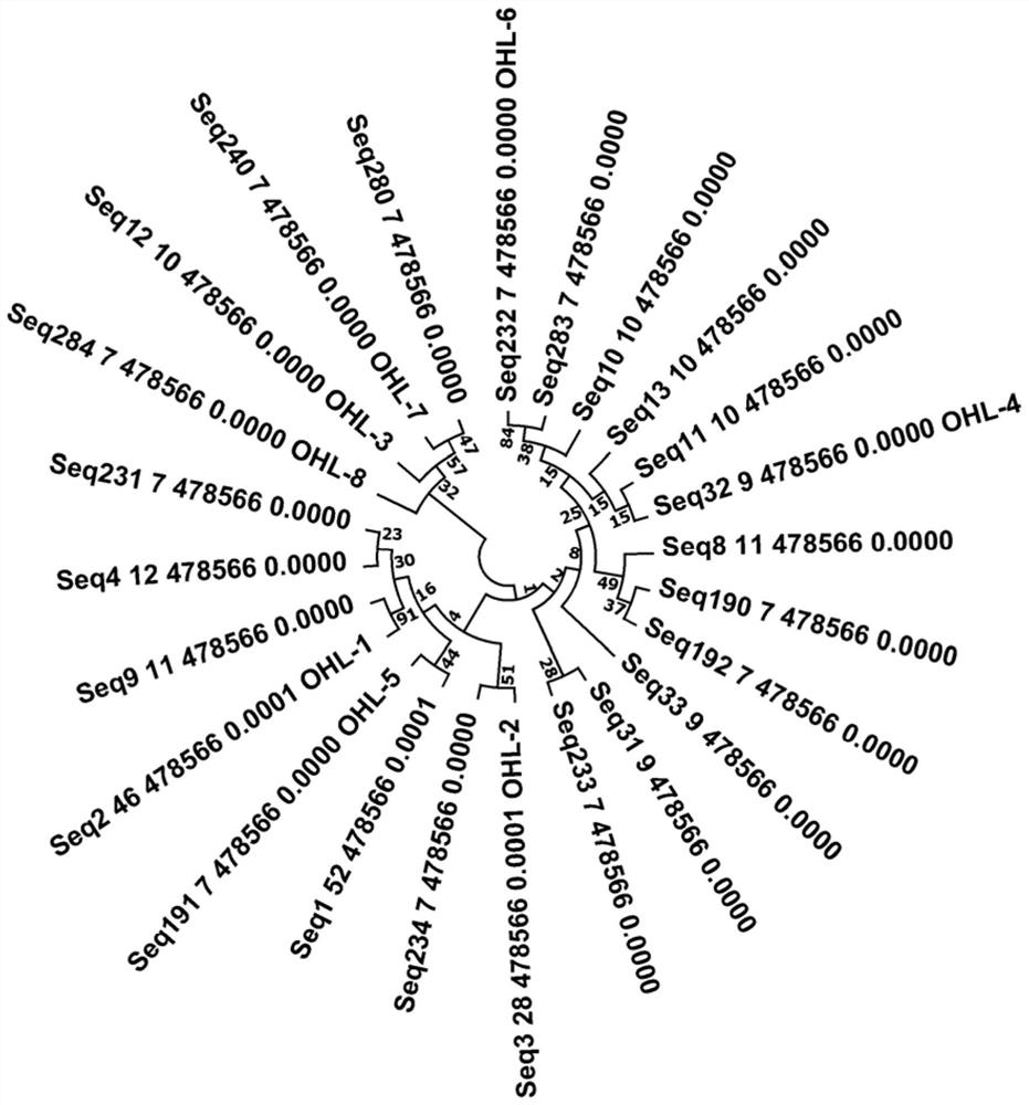
[0024]Based on Tissue-SELEX technology, the clinical rectal cancer tissue is a sieve sample, and the rectal cancer carcinoma is screened for negative sieve samples. First, the synthesis of the pro-group modification X-Aptamer library was synthesized, and then the initial library was incubated with rectal cancer carcinoma, isolated and retained and retained and retained by nucleic acid sequences bound to the cancer tissue as the first round of negative sieve libraries. At the same time, the non-combined nucleic acid sequence is incubated with clinical rectal cancer tissue, and then separated and retain the nucleic acid sequence combined with rectal cancer tissue as the first round of sieve library. Then, in the second round of screening, the positive sieve retained in the previous round is divided into three groups in sequential and sterilized water (blank control group), rectal cancer (experiment group), rec...
Embodiment 2
[0028]Example 2: Determining the strongest sequence of SW1463 cell line specific binding ability
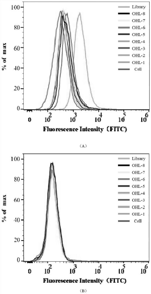
[0029]First, pretreatment of 8 candidate sequences and control library chains. The 8 nucleic acid aptamer sequences and the powder of the control library chain were centrifuged, and the sterile deionized water was diluted to a final concentration of 250 nm, degenerate in a metal bath in a 95 ° C, 10 min, and then placed Temperature temperature. SW1463 cells were simultaneously pretreated. SW1463 was washed twice with DPBS and 0.2% EDTA was added to 37 ° C. After washing with DPBS twice, cells were collected by wash buffer (WashingBuffer, 0.45% glucose, 5 mM magnesium chloride). After centrifugation, the binding buffer (BindingBuffer: DPBS, 0.45% glucose, 5 mM magnesium chloride, 100 mg / ltRNA, 1 g / lbsa) and after treatment were added and the derived nucleic acid body sequence and library. After mixing and mixing, it was placed at 4 ° C shaker for 1 hour. After the incubation, centrifug...
Embodiment 3
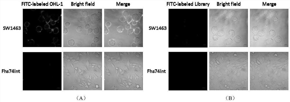
[0034]Example 3: Nucleic acid body OHL-1 combined with rectal cancer cells and weakness
[0035]The nucleic acid and body OHL-1 is further studied to further study the binding progents and strength of rectal cancer cells. The OHL-1 is first prepared. We set OHL-1 to different concentration gradients: 0 nm, 100 nm, 200 nm, 400 nm, 600 nm, 800 nm, 1000 nm, 1200 nm, 1400 nm, 1600 nm. High temperature denaturation at 95 ° C for 5 min, 10 min at 4 ° C for 10 min, and it was placed in normal temperature. Second, SW1463 cells were preprocessed. The original medium in the cell culture dish was discarded, and DPBS was added 2-3 times. Digestion 1min was digested with 0.2% EDTA to digest the SW1463 cells of the adhesive state from the culture dish, and then blew the cells by washing buffer (Washing Buffer, 5 mm magnesium chloride) and collected from the centrifuge tube. . Finally, oscillating mixing is placed at 4 ° C shaker incubation for 1 h. After incubation, then centrifuge with washing buff...
PUM
 Abstract
Description
Claims
Application Information
 - R&D Engineer
- R&D Manager
- IP Professional
- Industry Leading Data Capabilities
- Powerful AI technology
- Patent DNA Extraction
Browse by: Latest US Patents, China's latest patents, Technical Efficacy Thesaurus, Application Domain, Technology Topic, Popular Technical Reports.
© 2024 PatSnap. All rights reserved.Legal|Privacy policy|Modern Slavery Act Transparency Statement|Sitemap|About US| Contact US: help@patsnap.com


