
Intelligent automatic eye disease screening robot

A robot and eye disease technology, applied in ophthalmoscopes, eye testing equipment, medical science, etc., can solve problems such as difficult large-scale promotion, cumbersome process, and difficulty in fundus photography, so as to save manpower and material resources, speed up inspection, and benefit The effect of promotion

Active Publication Date: 2021-03-12
眼小医(温州)生物科技有限公司
View PDF9 Cites 1 Cited by
  • Summary
  • Abstract
  • Description
  • Claims
  • Application Information

AI Technical Summary

Problems solved by technology

[0002] Ophthalmic examination includes ocular surface examination, anterior segment examination and fundus examination. At present, common examination methods such as slit lamp microscope, front mirror, etc. are used to obtain information, and such ocular information of the examinee is obtained by taking pictures. Therefore, it is necessary Professional ophthalmologists use the autorefractor to obtain the initial diopter of the examinee, obtain the information of the patient's corneal curvature through the corneal topography examination, and then use the anterior segment camera and fundus camera to artificially focus on the cornea, iris and retina of the examinee's eye To obtain ocular surface, anterior segment and fundus photos, the whole process is relatively cumbersome, and it is difficult to promote it on a large scale in some areas with poor sanitation conditions, resulting in timely treatment of eye diseases
[0003] Patent CN107788945A discloses an ophthalmic inspection robot, which includes a head shell with an inspection mechanism inside. The inspection mechanism includes a head shell connection seat, a lighting device and a front camera device. The head shell connection seat is provided with a jaw rest movement The driving mechanism, front and rear focus driving mechanism, left and right movement driving mechanism and pupil horizontal rotation driving mechanism of the light spot, the present invention can replace experienced ophthalmologists to check vision and diopter through eye charts and test lenses, and through the front The camera device and the lighting source that can be rotated left and right to adjust the angle can capture the images of the outer eye and the anterior segment of the eyeball under a microscope similar to the slit lamp, and can transmit the inspection results and symptoms of the examinee to experienced ophthalmologists through the Internet in the form of images Or experts, professors consultation, but it is more difficult to photograph the fundus, and the eye position is not easy to align

Method used


Image

  • Intelligent automatic eye disease screening robot
  • Intelligent automatic eye disease screening robot
  • Intelligent automatic eye disease screening robot

Examples


Embodiment 1

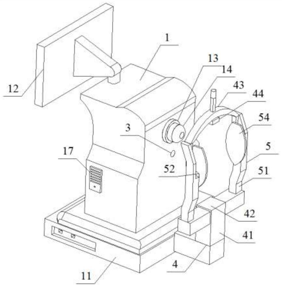
[0039] Such as figure 1As shown, an intelligent automatic eye disease screening robot includes a main body 1 for providing energy and data analysis, a positioning module 2 for automatic positioning of eyeballs, and a shooting module 3 for shooting faces and eyes. For fixing the head holder 4 of the patient's head,

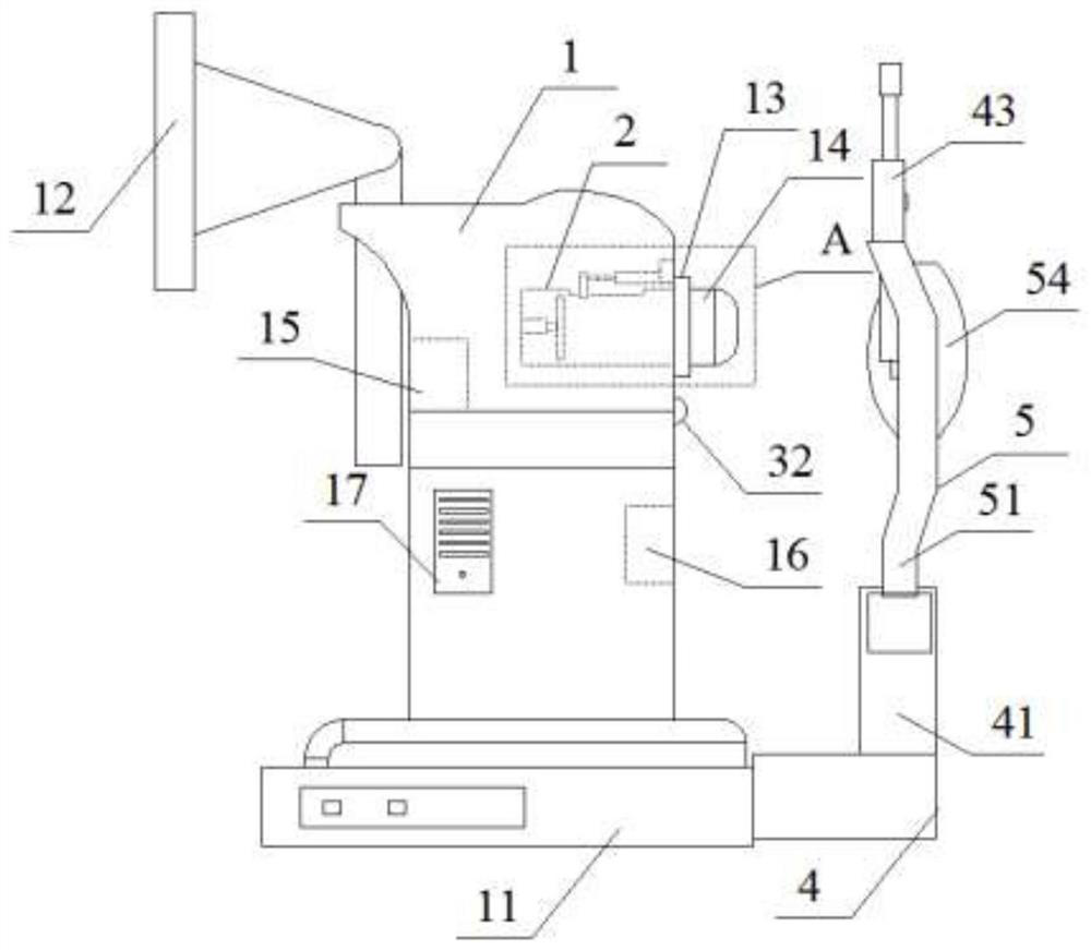
[0040] Such as figure 1 , 2 As shown, the bottom of the main body 1 is provided with a base 11, the front side of the main body 1 is provided with a display screen 12, the upper part of the rear side of the main body 1 is provided with an inspection slot 13, and a focusing tube 14 is embedded in the inspection slot 13, and the focusing tube 14 extends to Outside the main body 1, a groove 141 is opened on the upper part of the focusing tube 14 located inside the main body 1, and a data analysis module 15 for obtaining eye refraction data and corneal curvature data is provided inside the main body 1, and a storage module for storing data 16. One side of the base 1...

Embodiment 2

[0059] This embodiment is basically the same as Embodiment 1, except that a group of rotating motors 57 for controlling the rotation of the first connecting column 52 in the arc-shaped groove 511 are provided inside the clamping module 5;

[0060] Such as Figure 9-11 As shown, wherein, the bottom of the first connecting column 52 is provided with a slider 521, the bottom of the arc groove 511 is provided with a chute 513 for sliding the slider 521, the chute 513 is T-shaped, and the chute 513 is close to the patient's head The inside of one end is provided with a rotating motor 57, and the rotating motor 57 rotates along the slide rail 514 provided with the inside of the column 51. The output end of the rotating motor 57 is connected with a clip 571, and the upper end of the clip 571 is provided with a slider 521 for sliding. groove, the column 51 at the bottom of the slanting block 55 is provided with a groove for elongating and rotating the slanting block.

[0061] After t...

experiment example

[0063] The eye disease screening robot of the present invention was clinically tested, and 100 patients with conjunctivitis, 100 patients with keratitis, 100 patients with ametropia and 100 normal people were selected to use the eye disease screening robot of the present invention. Test, the test results are shown in the table below:

[0064] Table 1 Statistical table of examination results of various patients

[0065] Conjunctivitis Keratitis Refractive error normal 96 97 94 113

[0066] As can be seen from the test results in the above table, the eye disease screening robot of the present invention has a screening accuracy of 96% for patients with conjunctivitis, 97% for patients with keratitis, and 97% for patients with ametropia. The screening accuracy rate is 94%. It can be seen that the eye disease screening robot of the present invention can screen and diagnose eye diseases of patients with a low misdiagnosis rate, which can meet the needs of...


PUM


Abstract

The invention discloses an intelligent automatic eye disease screening robot which comprises a main body used for providing energy and data analysis, a positioning module used for automatically locating eyeballs, a shooting module used for face shooting and eye shooting and a head fixing frame used for fixing the head of a patient. A focusing cylinder is arranged on the rear upper part of the body; The positioning module is embedded in the focusing cylinder, the positioning module comprises a turntable used for rotating and switching the cameras and a positioning block used for locating the cameras, and the shooting module comprises an inner camera used for automatically shooting the ocular surface, the anterior segment of the eye and the fundus and an outer camera used for shooting the human face. Eyeballs of a patient can be automatically located, corneas, irises and retinas of the examined eye are focused, corresponding ocular surfaces, anterior segments and fundus photos are automatically shot; meanwhile, the corneal curvature and diopter of the examined eye are automatically obtained, professional ophthalmologists can be replaced to complete large-scale ophthalmologic comprehensive examination of the patient, and manpower and material resources are saved. The robot popularization is promoted.

Description

technical field [0001] The invention relates to the technical field of ophthalmic medical devices, in particular to an intelligent automatic eye disease screening robot. Background technique [0002] Ophthalmic examination includes ocular surface examination, anterior segment examination and fundus examination. At present, common examination methods such as slit lamp microscope, front mirror, etc. are used to obtain information, and such ocular information of the examinee is obtained by taking pictures. Therefore, it is necessary Professional ophthalmologists use the autorefractor to obtain the initial diopter of the examinee, obtain the information of the patient's corneal curvature through the corneal topography examination, and then use the anterior segment camera and fundus camera to artificially focus on the cornea, iris and retina of the examinee's eye To obtain ocular surface, anterior segment and fundus photos, the whole process is relatively cumbersome, and it is di...

Claims


Application Information

Patent Timeline
IPC IPC(8): A61B3/14A61B3/00A61B3/12A61B3/032A61B3/107
CPCA61B3/14A61B3/0008A61B3/0016A61B3/0083A61B3/12A61B3/032A61B3/107
Inventor 陈蔚李中文郑钦象
Owner 眼小医(温州)生物科技有限公司
Learn More