
Method for preparing cell microvesicles based on shear stress stimulation

A technology of stimulation and shear stress, applied in the field of bioengineering, can solve the problems of lack of specificity of EVs, uneven concentration of EVs, and easy pollution in the operation process, so as to solve the problems of long time consumption, reduce sample processing steps, and good stability Effect

Pending Publication Date: 2021-03-09
CHONGQING UNIV
View PDF4 Cites 2 Cited by
  • Summary
  • Abstract
  • Description
  • Claims
  • Application Information

AI Technical Summary

Problems solved by technology

[0004] The conventional centrifugation method is the most widely used, but there are many disadvantages, such as high cost of reagents, long processing time, cumbersome process, and easy contamination during operation, etc.
Moreover, it lacks specificity for EVs, is prone to produce heterogeneous polymer particles, and has problems such as uneven concentration of extracted EVs.

Method used


Image

  • Method for preparing cell microvesicles based on shear stress stimulation
  • Method for preparing cell microvesicles based on shear stress stimulation
  • Method for preparing cell microvesicles based on shear stress stimulation

Examples


Embodiment 1

[0036] The method for preparing cell microvesicles based on shear stress stimulation comprises the following steps:

[0037] 1) Add the macrophages extracted from the RAW 264.7 cell line to a 50mL culture flask, and add 3mL of DMEM medium and 10% normal serum to the culture flask, and place it at 37°C Culture in an incubator. After the cells proliferate to 90% of the total volume of the entire culture flask, the culture is terminated. At this time, the cells in the culture flask are about 1×10 8 indivual;

[0038] 2) Next, place the cells on an orbital shaker and culture them for 4 hours at a rotational speed of 50r / min (i.e., undergo shear stress treatment), scrape the cells from the culture flask with a plastic cell scraper, and quickly blow them with a pipette to fully absorb the cells. Disperse into a 10mL centrifuge tube, vortex for 30s, centrifuge at 1000rmp for 5min, take the supernatant into a new centrifuge tube, centrifuge again at 10000rmp for 10min, remove the sup...

Embodiment 2

[0042] The method for preparing cell microvesicles based on shear stress stimulation comprises the following steps:

[0043] 1) Add the macrophages extracted from the RAW 264.7 cell line to a 50mL culture flask, and add 3mL of DMEM medium and 10% normal serum to the culture flask, and place it at 37°C Culture in an incubator. After the cells proliferate to 90% of the total volume of the entire culture flask, the culture is terminated. At this time, the cells in the culture flask are about 1×10 8 indivual;

[0044]2) Next, place the cells on an orbital shaker and culture them at a speed of 70r / min for 2 hours (i.e., undergo shear stress treatment), scrape the cells from the culture flask with a plastic cell scraper, and quickly blow them with a pipette to fully absorb the cells. Disperse into a 10mL centrifuge tube, vortex for 30s, centrifuge at 1000rmp for 5min, take the supernatant into a new centrifuge tube, centrifuge again at 10000rmp for 10min, remove the supernatant, an...


PUM

PropertyMeasurementUnit
diameteraaaaaaaaaa
diameteraaaaaaaaaa
particle sizeaaaaaaaaaa

Abstract

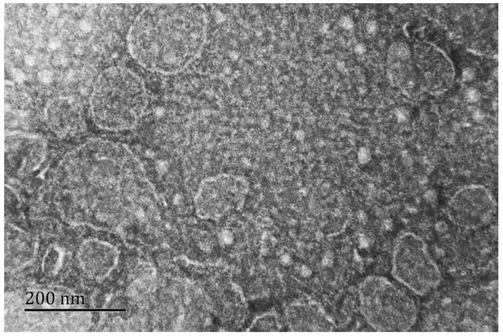
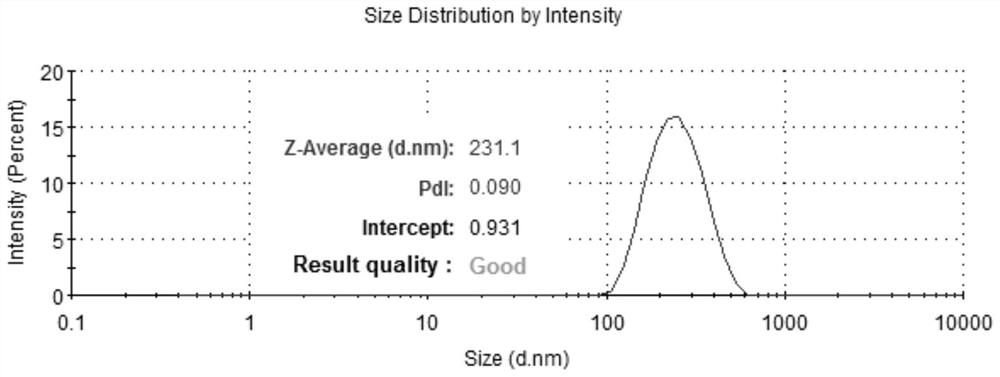
The invention discloses a method for preparing cell microvesicles based on shear stress stimulation, and belongs to the technical field of bioengineering. The method comprises the following preparation steps of: taking cells, adding the cells into a culture solution, culturing, carrying out shear stress treatment after the cells grow to 90%, collecting the cells, whirling, centrifuging, taking supernatant, and centrifuging again to obtain precipitate; and putting the precipitate into phosphate buffered saline, blowing, beating, uniformly mixing, centrifuging, taking supernatant, centrifuging again to obtain secondary precipitate, adding water, carrying out hypotonic treatment, and centrifuging to obtain the cell microvesicles. According to the method for preparing the cell microvesicles based on shear stress stimulation, EVs with high content can be conveniently and quickly obtained; meanwhile, in the preparation process, chemical reagent stimulation is not needed, heterogeneous polymer particles cannot be generated, and manpower, material resources and financial resources are saved; and the prepared cell microvesicles are uniform in size, the average particle size is 231nm, and the method has popularization and application values in the field of biological materials.

Description

technical field [0001] The invention relates to the technical field of bioengineering, in particular to a method for preparing cell microvesicles under shear stress stimulation. Background technique [0002] Cell microvesicles (Extracellular Vesicles, EVs) are mostly 100-1000nm in diameter and are formed by cell membrane budding. Cells respond to cell activation, pH changes, hypoxia, radiation, damage or cell stress by releasing EVs. Pathologically, microvesicle budding has also been suggested as a mechanism for repairing damaged plasma membrane after plasma membrane injury. EVs can be found in many body fluids, including blood, urine, tears, and saliva, and participate in various physiological and pathological processes, such as cardiovascular diseases. EVs are now increasingly recognized as important carriers of circulating markers for intercellular communication, disease diagnosis, and prognosis. However, the amount of EVs produced by the cells itself is not large, and ...

Claims


Application Information

Patent Timeline
Patent Type & Authority Applications(China)
IPC IPC(8): C12N5/0786A61K47/46
CPCC12N5/0645A61K47/46C12N2509/10
Inventor 王贵学王良婷王溢吴伟
Owner CHONGQING UNIV
Learn More