Epilepsy focus positioning method and system
A positioning method and lesion technology, applied in the field of image processing, can solve problems such as error-prone and large differences in epilepsy, and achieve the effects of saving running time, solving incomplete features, and improving sensitivity and specificity
- Summary
- Abstract
- Description
- Claims
- Application Information
AI Technical Summary
Problems solved by technology
Method used
Image
Examples
Embodiment 1
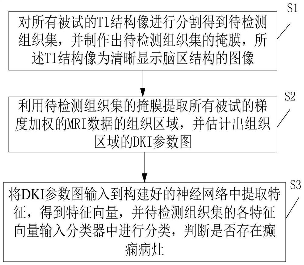
[0040] The embodiment of the present invention provides a method for locating epileptic focus, which is applied in conventional MRI-negative epilepsy, and the patient's condition can be effectively controlled through timely and accurate focus locating diagnosis. Such as figure 1 shown, including the following steps:
[0041] Step S1: Segment the T1 structural images of all subjects to obtain the tissue set to be detected, and make a mask of the tissue set to be detected. The T1 structural image is an image that clearly shows the structure of the brain region.
[0042] The process of realizing the above step S1 in the embodiment of the present invention is as follows:
[0043] Step S11: Convert all subjects' T1 structural images from two-dimensional images to three-dimensional images; in practical applications, dcm2niigui software can be used to convert the format of all subjects' T1 structural images from Dicom to 3Dnifty.
[0044] Step S12: Preprocessing the 3D image, for e...
Embodiment 2
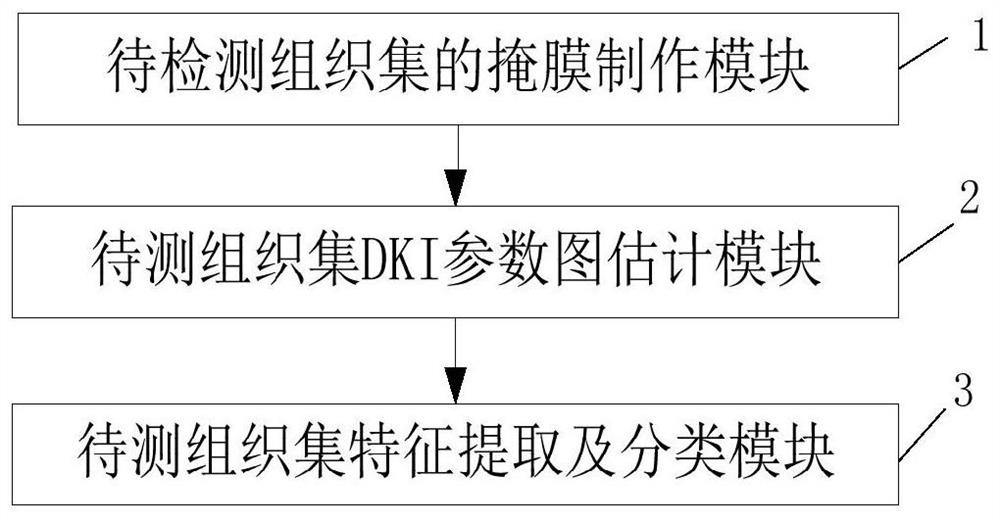
[0067] An embodiment of the present invention provides an epileptic focus positioning system, such as image 3 shown, including:
[0068] The mask production module 1 of the tissue set to be detected is used to segment the T1 structural images of all subjects to obtain the tissue set to be detected, and to make a mask of the tissue set to be detected. The T1 structural image clearly shows the brain region The image of the structure; this module executes the method described in step S1 in Embodiment 1, which will not be repeated here.
[0069] The DKI parameter map estimation module 2 of the tissue set to be tested is used to extract the tissue area of the gradient-weighted MRI data of all subjects by using the mask of the tissue set to be tested, and estimate the DKI parameter map of the tissue area; this module implements The method described in step S2 in Example 1 will not be repeated here.
[0070] The feature extraction and classification module 3 of the tissue set to...
Embodiment 3
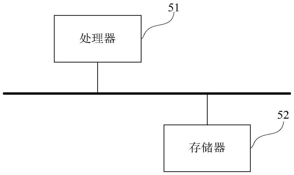
[0073] An embodiment of the present invention provides a computer device, such as image 3 As shown, the device may include a processor 51 and a memory 52, wherein the processor 51 and the memory 52 may be connected via a bus or in other ways, image 3 Take connection via bus as an example.
[0074] The processor 51 may be a central processing unit (Central Processing Unit, CPU). Processor 51 can also be other general processors, digital signal processor (Digital Signal Processor, DSP), application specific integrated circuit (Application Specific Integrated Circuit, ASIC), field programmable gate array (Field-Programmable Gate Array, FPGA) or Other chips such as programmable logic devices, discrete gate or transistor logic devices, discrete hardware components, or combinations of the above-mentioned types of chips.
[0075] As a non-transitory computer-readable storage medium, the memory 52 can be used to store non-transitory software programs, non-transitory computer-execu...
PUM
 Abstract
Description
Claims
Application Information
 - Generate Ideas
- Intellectual Property
- Life Sciences
- Materials
- Tech Scout
- Unparalleled Data Quality
- Higher Quality Content
- 60% Fewer Hallucinations
Browse by: Latest US Patents, China's latest patents, Technical Efficacy Thesaurus, Application Domain, Technology Topic, Popular Technical Reports.
© 2025 PatSnap. All rights reserved.Legal|Privacy policy|Modern Slavery Act Transparency Statement|Sitemap|About US| Contact US: help@patsnap.com


