
Method for preparing monoclonal antibody through rapid immunization

A monoclonal antibody and rapid immunization technology, applied in the field of IVD diagnosis, can solve the problems of cumbersome and time-consuming operations, affecting antibody titers, and long immunization cycles, and achieve simple immunization operations, increased antibody titers, and low antigen usage Effect

Inactive Publication Date: 2021-01-05
巴德生物科技有限公司
View PDF5 Cites 0 Cited by
  • Summary
  • Abstract
  • Description
  • Claims
  • Application Information

AI Technical Summary

Problems solved by technology

[0002] At present, the preparation of hybridoma monoclonal antibody is mainly based on cell fusion technology to fuse B lymphocytes secreting specific antibodies with bone marrow cells with unlimited proliferation ability to form specific antibodies against an antigenic epitope. This process requires immunization. Currently, commonly used immunization methods include intraperitoneal injection, intramuscular injection and subcutaneous injection. These three immunizations are usually repeated 3-4 times at intervals of 14 days. It is estimated that it will take 49-63 days for the immunization to reach fusion conditions. The immunization cycle is long, and adjuvant emulsification is required, and the operation is cumbersome and time-consuming; this method has certain defects, such as: 1, after the antigen injection, the spleen is not fully stimulated, and part of it is decomposed, which affects the antibody titer; 2, the existing technology During the emulsification process, whether using manual push or phacoemulsification, there is excessive heat during the operation process, which leads to antigen denaturation

Method used


Image

  • Method for preparing monoclonal antibody through rapid immunization

Examples


Embodiment Construction

[0028] The following will clearly and completely describe the technical solutions in the embodiments of the present invention in conjunction with the accompanying drawings in the embodiments of the present invention; obviously, the described embodiments are only part of the embodiments of the present invention, not all embodiments, based on The embodiments of the present invention and all other embodiments obtained by persons of ordinary skill in the art without making creative efforts belong to the protection scope of the present invention.

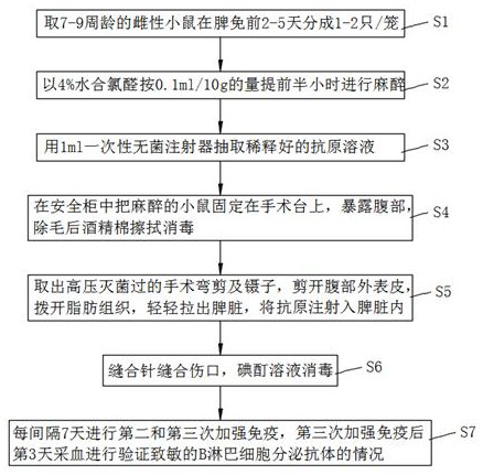
[0029] The present invention provides a technical solution: please refer to figure 1 , a method for preparing monoclonal antibody rapid immunization, comprising the following steps:

[0030] Step 1. Among the cultured female mice, 7-9 week-old female mice are taken and divided into cages, with 1-2 mice in each cage.

[0031] Step 2: Anesthetize the caged female mice with 4% chloral hydrate in an amount of 0.1ml / 10g.

[0032] Step 3: Dr...


PUM


Abstract

The invention discloses a method for preparing a monoclonal antibody through rapid immunization, which belongs to the technical field of diagnosis, and comprises the following steps: S1, taking animals which are not less than 7 weeks old from cultured animals for cage separation, S2, anesthetizing the animals by using 4% chloral hydrate according to the amount of 0.1 ml / 10g, S3, extracting a diluted antigen solution by using a 1ml disposable sterile syringe, and S4, fixing the narcotized animal on an operating table in a clean environment, exposing the abdomen, and removing hairs from the abdomen; S5, cutting off the outer epidermis of the abdomen by using a pair of surgical curved scissors, pushing aside adipose tissues, slightly pulling out the spleen by using tweezers, and injecting anantigen into the spleen; S6, suturing the wound by using a suture needle; S7, carrying out second and third immunization every 3-14 days. Blood is collected 25 days after the third immunization to verify the antibody secretion condition of the sensitized B lymphocytes; the invention has the advantages of reasonable and practical design, low antigen usage amount, reduced cost, improved antibody titer, and elimination of false positive to some extent. The immune operation is simple, and the situation of unsuccessful immunization caused by immunization to the subcutaneous part does not need to beworried.

Description

technical field [0001] The invention relates to the technical field of IVD diagnosis, in particular to a method for preparing monoclonal antibodies for rapid immunization. Background technique [0002] At present, the preparation of hybridoma monoclonal antibody is mainly based on cell fusion technology to fuse B lymphocytes secreting specific antibodies with bone marrow cells with unlimited proliferation ability to form specific antibodies against an antigenic epitope. This process requires immunization. Currently, commonly used immunization methods include intraperitoneal injection, intramuscular injection and subcutaneous injection. These three immunizations are usually repeated 3-4 times at intervals of 14 days. It is estimated that it will take 49-63 days for the immunization to reach fusion conditions. The immunization cycle is long, and adjuvant emulsification is required, and the operation is cumbersome and time-consuming; this method has certain defects, such as: 1,...

Claims


Application Information

Patent Timeline
IPC IPC(8): C07K16/06
CPCC07K16/00C07K2317/10
Inventor 邢贞华李博
Owner 巴德生物科技有限公司
Learn More