Automatic analysis method for hemodynamic parameters of ocular capillaries

An analysis method and eye microvascular technology, applied in image analysis, image data processing, image enhancement, etc., can solve problems such as huge workload, limited blood vessels, discontinuous blood vessels, etc., to reduce human-computer interaction, improve accuracy, and process fast effect

Active Publication Date: 2021-01-01
美十光学科技(广东)有限公司
View PDF8 Cites 2 Cited by
  • Summary
  • Abstract
  • Description
  • Claims
  • Application Information

AI Technical Summary

Problems solved by technology

Domestic scientific research institutions have scattered eye imaging research technology, and most of the research and development equipment is to track and imitate the previous generation of foreign technology products, and there are defects such as imperfect supporting analysis software, batch image processing and intelligent analysis that are far from meeting scientific research and clinical needs.
[0003] The existing eye microvessel acquisition system is realized by integrating digital image acquisition devices such as high-speed camera and video acquisition card into the standard slit lamp platform. During the acquisition process, slight shaking of the patient will cause a large shift in the image, so the entire video The images that can be used for blood flow analysis in the image only account for less than 20% of the entire video image. The existing system uses manual annotation by doctors, and the workload is very huge. In addition, the blood vessels displayed in the eye microvascular images are discontinuous. There are discontinuities between blood vessels in a single image, and the blood vessels used for analysis in a single image obtained by traditional methods are very limited, which greatly reduces the availability of blood vessels; Using manual methods will cause different errors due to different individuals or different degrees of fatigue, and at the same time it is extremely time-consuming

Method used


Image

  • Automatic analysis method for hemodynamic parameters of ocular capillaries
  • Automatic analysis method for hemodynamic parameters of ocular capillaries

Examples


Embodiment Construction

[0042] The following description serves to disclose the present invention to enable those skilled in the art to carry out the present invention. The preferred embodiments described below are only examples, and those skilled in the art can devise other obvious variations.

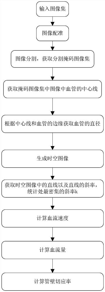
[0043] An automatic analysis method for eye microvascular hemodynamic parameters, the dynamic parameters mainly include blood flow velocity, blood flow and vessel wall shear rate, specifically comprising the following steps:

[0044] Step 1: Input the image set I in the video file that needs to analyze the hemodynamic parameters of eye microvessels o , and enter the relevant parameters.

[0045] The video file is obtained by shooting eye microvessels with a camera or the like. For the image set I o , the video file is composed of many consecutive images, the image set I o It's a collection of these images.

[0046] Step 2: To image collection I o Image registration is performed on the continuous images...


PUM


Abstract

The invention provides an automatic analysis method for hemodynamic parameters of ocular capillaries. The method comprises the following steps: 1, inputting an image set Io which needs to be subjectedto analysis of hemodynamic parameters of ocular capillaries in a video file containing eye microvessel information; 2, performing image registration on continuous images in the image set Io; 3, performing image segmentation on the registered image to obtain a segmentation mask image set Sr of the eye microvessels; 4, obtaining a center line of an eye microvessel in the segmentation mask image setSr; 5, fitting the center line, extracting the edge of the ocular capillaries in the segmentation mask image, and calculating the average diameter, the inner diameter and the outer diameter of the ocular capillaries; 6, forming a space-time image; 7, carrying out histogram equalization processing on the obtained space-time image; 8, performing straight line detection processing on the space-timeimage, performing statistics on the slopes of the obtained straight lines, and obtaining the slope k with the densest distribution; 9, calculating the blood flow velocity Va of the ocular capillaries;and 10, calculating the blood flow and the tube wall stress cutting rate.

Description

technical field [0001] The invention relates to the field of parameter analysis of eye microvessels, in particular to an automatic analysis method for hemodynamic parameters of eye microvessels. Background technique [0002] The eyeball and the brain share the blood supply of the internal carotid artery. Eye microvascular function is a potential indicator of ocular surface inflammation and systemic microvascular disease. effects etc. provide an important basis. With the rapid development of ophthalmic imaging technology in recent years, the advent and development of new imaging technologies such as functional imaging OCT and ocular microvascular function analysis provide important information for the diagnosis and treatment of ophthalmic diseases, and its morphological measurement function has been applied to ocular tissue In the precise analysis of function, it is called a milestone achievement of ophthalmology diagnostic technology. Foreign developed countries have inves...

Claims


Application Information

Patent Timeline
Patent Type & Authority Applications(China)
IPC IPC(8): G06T7/00G06T7/13G06T5/40
CPCG06T7/0012G06T7/13G06T5/40G06T2207/10016G06T2207/30101G06T2207/30041
Inventor 袁进王耿媛贠照强肖鹏段铮昱骆仲舟黄远聪
Owner 美十光学科技(广东)有限公司
Why Eureka
Try Eureka