
Cell block collection device and cell block collection method

A collection device and collection method technology, applied in the direction of measuring devices, sampling, instruments, etc., can solve the problems of wrong or missed detection, unfavorable microscopic inspection, and affecting the accuracy of exfoliated cells, etc.

Active Publication Date: 2020-10-23
郑志森 +1
View PDF9 Cites 0 Cited by
  • Summary
  • Abstract
  • Description
  • Claims
  • Application Information

AI Technical Summary

Problems solved by technology

Although this method can fix the exfoliated cells within a certain range, the enrichment effect of the cells is not good, especially the exfoliated cells in the exfoliated cell liquid collected at the beginning are not much, and the exfoliated cells with lesions will be less , when cell sectioning is performed, the obtained test sample may happen to not contain diseased exfoliated cells, which leads to the following: firstly, it is not conducive to the later stage of microscopic examination and pathological examination, resulting in inaccurate test results; secondly, if you want to To make the detection results more accurate, it is necessary to do more microscopic examination and detection of the same sample, resulting in a cumbersome detection process and high detection costs; Acquisition and enrichment of target cells of exfoliated cells in fluid and exfoliated cells in sputum), which in turn affects the accuracy of detection of exfoliated cells in the later stage
[0006] For the current various tumor detection, when the tumor is in the early stage and has not yet become cancerous, the amount of tumor cells in the form of exfoliated cells in the body fluid is relatively small, so the conventional method of collecting exfoliated cells is used to obtain In the sample, the amount of the target cells to be tested is very small, resulting in the final test result showing that the cell sample does not contain diseased cells, which is easy to cause false detection or missed detection.

Method used


Image

  • Cell block collection device and cell block collection method
  • Cell block collection device and cell block collection method
  • Cell block collection device and cell block collection method

Examples


preparation example

[0043] Preparation example Gelatin preparation: To prepare an aqueous solution of gelatin, add agar, tissue fixative and water at a mass ratio of 1:1:8, and then stir evenly. Wherein, the tissue fixative is medical formaldehyde or medical alcohol; the medical formaldehyde is a formaldehyde aqueous solution with a formaldehyde mass fraction of 35-40%, and the medical alcohol is an ethanol aqueous solution with a volume fraction of 75%. The tissue fixative selected in this preparation example is an aqueous formaldehyde solution with a mass fraction of formaldehyde of 38%.

Embodiment 1

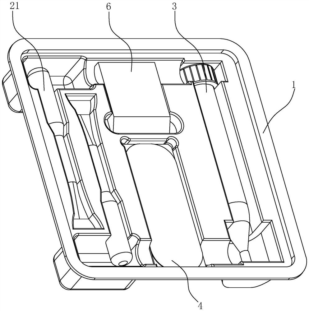
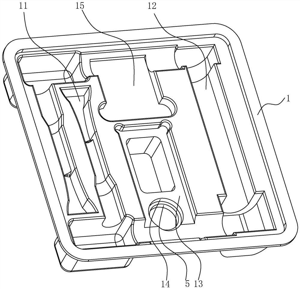
[0047] A cell block collection device, such as figure 1 and figure 2 As shown, it includes a support plate 1 and several auxiliary parts. The support plate 1 is made of plastic and has certain deformation capabilities. The support plate 1 is provided with grooves of different shapes. The opening shape of each groove and the corresponding The shape of the auxiliary parts is adapted so that each auxiliary part can be securely snapped into the corresponding groove. The auxiliary parts include auxiliary material tubes, a fixed support plate 4, a special centrifuge tube 3 for containing exfoliated cell liquid during centrifugation, a limiter 5 and a tissue embedding box 6. The auxiliary material tube includes a sealed cell fixing glue tube 21 with built-in cell fixing glue.

[0048] Such as figure 2 As shown, the grooves include auxiliary material tube grooves 11 , centrifuge tube grooves 12 , pallet grooves 13 , stopper grooves 14 and embedding cassette grooves 15 . Specific...

Embodiment 2

[0067] The difference between this embodiment and embodiment 1 is that the operating process parameters are different, specifically:

[0068] In step S1, after the obtained body fluid is pretreated to obtain exfoliated cell fluid with less impurity content, the specific steps of the pretreatment are as follows: 1. Take a clean common centrifuge tube with a volume of 50 mL, and take the obtained body fluid Pour 20mL into an ordinary centrifuge tube, then centrifuge at 3000rpm for 5min. Other operations of step S1 are the same as those in Embodiment 1.

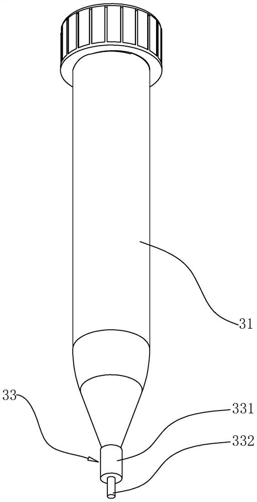
[0069] In step S2, the special centrifuge tube 3 containing the treated exfoliated cell liquid is placed in the centrifuge, centrifuged at a speed of 3000rpm for 5 minutes, and then the supernatant is poured off, and the exfoliated cells are collected in the straight tube 32; step S3-S6 is the same as embodiment 1.


PUM

PropertyMeasurementUnit
pore sizeaaaaaaaaaa

Abstract

The invention discloses a cell block collection device and a cell block collection method, and belongs to the technical field of collection of cells in body fluid. The technical problem of low detection rate when cancer detection is carried out by adopting cell slices when the content of cancerous cells in the body fluid is relatively low is solved. The cell collection device comprises a supporting plate, a plurality of grooves are formed in the supporting plate, and an auxiliary material tube, a fixed supporting plate, a special centrifugal tube, a tissue embedding box and embedding paper areclamped in the grooves respectively. A use method of the cell collection device comprises the following steps: preparing raw materials; centrifugally filtering exfoliated cell sap; performing cell precipitation hardening; fixing exfoliated cell blocks; and embedding and paraffinizing the exfoliated cell blocks. The cell collection device can be used for collecting and preparing cell blocks of cast-off cells in samples such as pleural effusion, ascites, urine and sputum, has the advantages of efficient enrichment of a small number of cast-off cells and unsupported preparation of the cell blocks, facilitates later cell slicing, and improves the detection rate of later pathological detection.

Description

technical field [0001] The present application relates to the technical field of cell collection in body fluids, and more specifically, it relates to a cell block collection device and a cell block collection method. Background technique [0002] In the medical field, for the early diagnosis of lung cancer and liver cancer, at present, chest X-ray and chest CT scan are mainly used for initial diagnosis, followed by the acquisition, preparation and detection of tissue slices. However, the acquisition of tissue slices is often a damaged slice sample acquisition method, and the relevant tissue cells can only be obtained after wounds are made on the corresponding parts of the human body. Therefore, the subsequently developed non-destructive cell slice detection technology has been widely used by everyone. Non-destructive cell slicing is to extract the exfoliated cells of the target diagnostic site, and then slice and detect the extracted exfoliated cells after further processin...

Claims


Application Information

Patent Timeline
Patent Type & Authority Applications(China)
IPC IPC(8): G01N1/28G01N1/34G01N1/36
CPCG01N1/286G01N1/34G01N1/36G01N2001/2873Y02E60/10
Inventor 郑志森江鑫烨陈明林双平
Owner 郑志森
Learn More