Segmentation method, device and terminal for epithelial cell nucleus in prostate cancer pathological image

A pathological image, prostate cancer technology, applied in image analysis, image enhancement, image data processing and other directions, to achieve the effect of improving the accuracy of judgment

Active Publication Date: 2020-07-10
SUN YAT SEN MEMORIAL HOSPITAL SUN YAT SEN UNIV +1
View PDF3 Cites 7 Cited by
  • Summary
  • Abstract
  • Description
  • Claims
  • Application Information

AI Technical Summary

Problems solved by technology

However, since both epithelial cell nuclei and stromal cell nuclei contain nucleic acid substances, there is no significant difference in color after being specifically combined with a stain, and it is difficult to classify epithelial cell nuclei and stromal cell nuclei using traditional image segmentation algorithms
In addition, the adjacency and overlapping phenomena between nuclei make the accuracy of nucleus segmentation still have a lot of room for improvement.

Method used


Image

  • Segmentation method, device and terminal for epithelial cell nucleus in prostate cancer pathological image
  • Segmentation method, device and terminal for epithelial cell nucleus in prostate cancer pathological image
  • Segmentation method, device and terminal for epithelial cell nucleus in prostate cancer pathological image

Examples


Embodiment 1

[0059] Please refer to figure 1 , this embodiment proposes a method for segmenting epithelial cell nuclei in pathological images of prostate cancer, which can be applied to the processing of pathological images of prostate cancer, especially including analyzing the shape, texture and color of epithelial cell nuclei in pathological images, and then realizing the segmentation Efficient prediction of prostate cancer.

[0060] The method for segmenting epithelial cell nuclei in the pathological image of prostate cancer will be described in detail below.

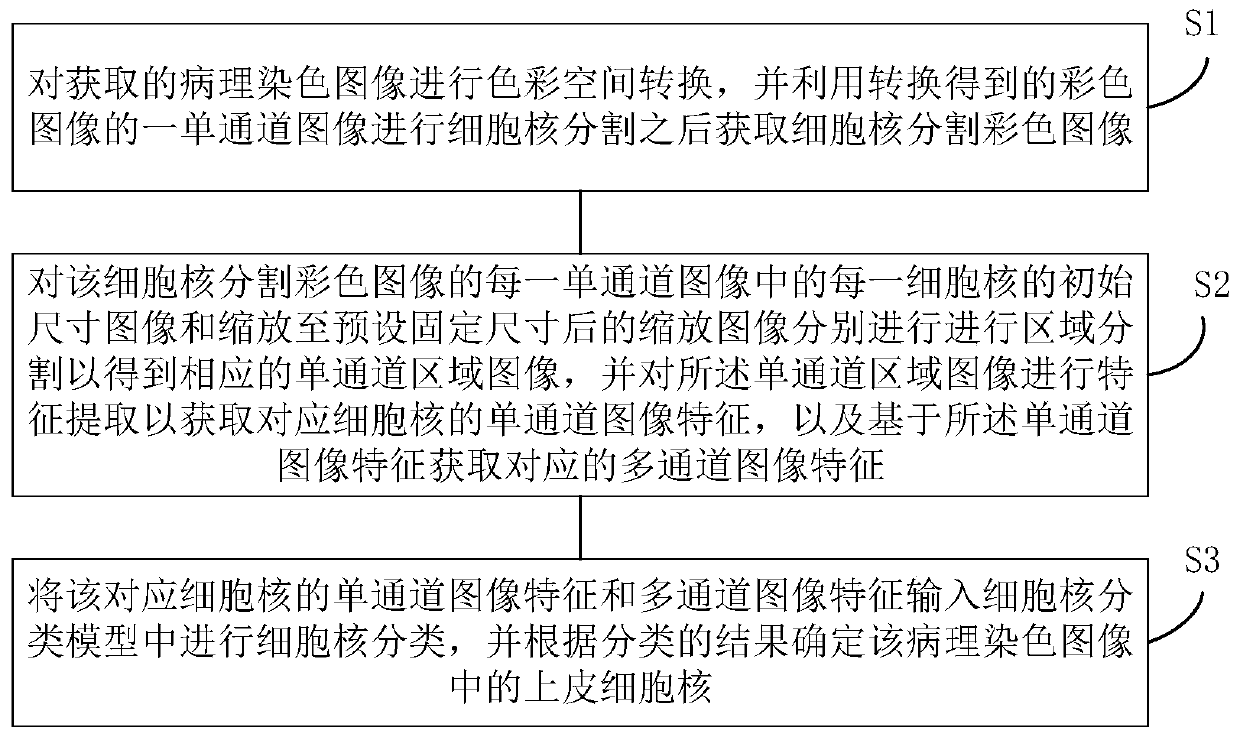
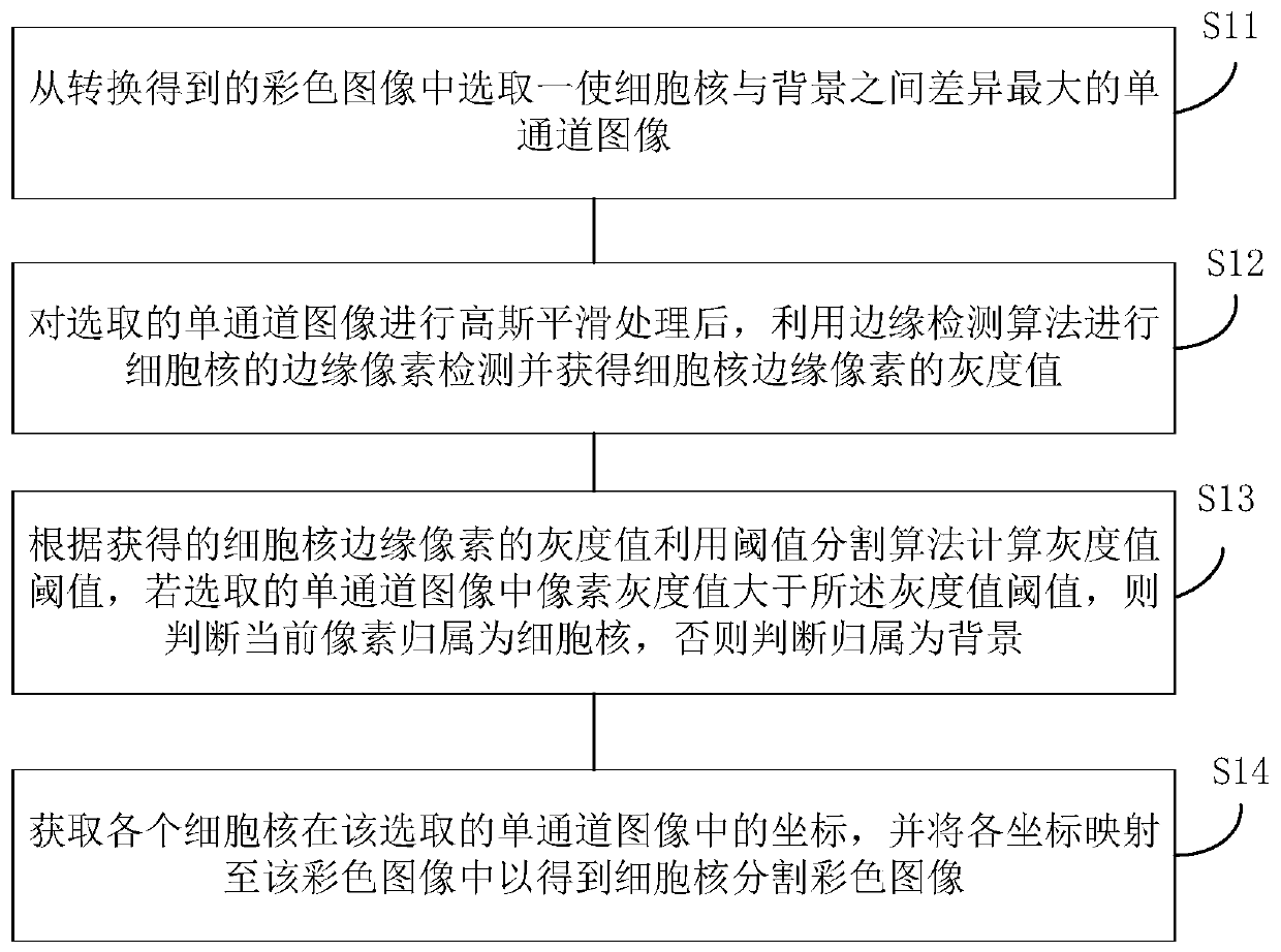
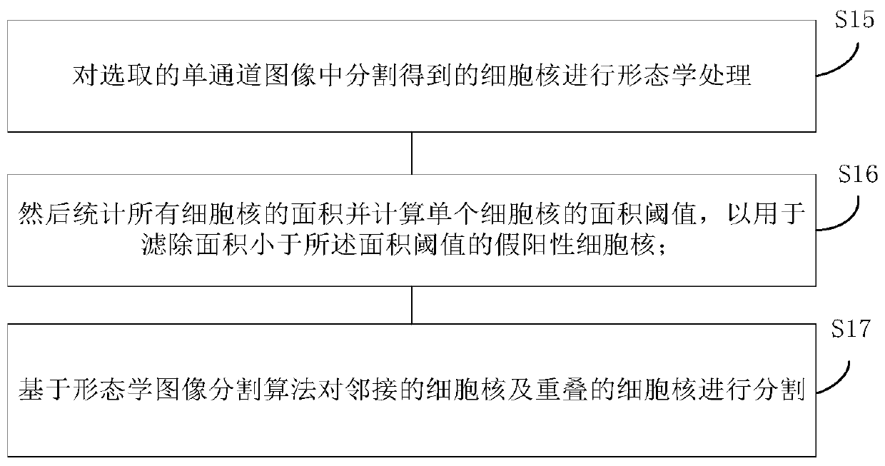
[0061] Step S1 , performing color space conversion on the obtained pathologically stained image, and using a single-channel image of the converted color image to perform cell nucleus segmentation to obtain a cell nucleus segmented color image.

[0062] This step S1 is mainly used to segment the nucleus in the prostate body, that is, to identify the nucleus in the pathologically stained image. In this embodiment, considering tha...

Embodiment 2

[0139] Please refer to Figure 8 , based on the method of the above-mentioned embodiment 1, this embodiment proposes a device 10 for segmenting epithelial cell nuclei in a pathological image of prostate cancer, including:

[0140] A cell nucleus segmentation module 110, configured to perform color space conversion on the acquired pathological staining image, and obtain a cell nucleus segmentation color image after performing cell nucleus segmentation based on a single-channel image of the converted color image;

[0141] The cell nucleus feature extraction module 120 is used to segment the initial size image of each cell nucleus in each single-channel image of the cell nucleus segmentation color image and the zoomed image after zooming to a preset fixed size to obtain corresponding single-channel images. channel region image, and performing feature extraction on the single-channel region image to obtain single-channel image features corresponding to the nucleus, and obtain corr...


PUM


Abstract

The embodiment of the invention discloses a segmentation method, device and terminal for epithelial cell nucleuses in a prostate cancer pathological image, and the method comprises the steps: carryingout the color space conversion of an obtained pathological staining image, and carrying out the cell nucleus segmentation based on a single-channel image of a converted color image; performing regionsegmentation on the initial size image and the scaled image of each cell nucleus in each single-channel image of the cell nucleus segmentation color image to obtain a single-channel region image, andperforming feature extraction on each single-channel region image; inputting the obtained single-channel image features and multi-channel image features of the cell nucleus into a cell nucleus classification model for cell nucleus classification, and determining epithelial cell nucleuses in the pathological staining image according to a classification result. According to the technical scheme, the problem that epithelial cell nucleuses in the prostate are difficult to accurately segment in the prior art can be well solved, so the accuracy of judgment on pathological diagnosis, severity and the like of the prostate cancer is improved.

Description

technical field [0001] The present invention relates to the technical field of pathological image processing, in particular to a method, device and terminal for segmenting epithelial cell nuclei in pathological images of prostate cancer. Background technique [0002] The prostate is an important organ of the male genitourinary system. A normal prostate gland is composed of lumens and epithelial cells surrounding the lumens. The area between the glands is composed of stroma and stromal cells. Prostate cancer mainly occurs in epithelial cells. The malignant expansion of epithelial cells will cause the cavity of the prostate to shrink or even be completely blocked, seriously affecting various functions of the prostate, seriously threatening the health and quality of life of men, and even the lives of patients. [0003] Prostate cancer is a malignant tumor of epithelial origin. In its clinical diagnosis, pathological diagnosis is the "gold standard", and pathological images play...

Claims


Application Information

Patent Timeline
Patent Type & Authority Applications(China)
IPC IPC(8): G06T7/11G06T7/136G06T7/45G06T7/62G06T7/90G06K9/62G06K9/46
CPCG06T7/11G06T7/90G06T7/62G06T7/45G06T7/136G06T2207/10056G06T2207/30024G06T2207/30096G06V10/50G06F18/24Y02A90/10
Inventor 赖义明郭正辉彭圣萌吴宛桦范辉阳雷震李嘉路华芮张游龙李骁
Owner SUN YAT SEN MEMORIAL HOSPITAL SUN YAT SEN UNIV
Why Eureka
Try Eureka