


Quick Research
Generate reliable direction feasibility study reports for your R&D in just a few steps.

Technical Q&A
Discover and master advanced knowledge NOW. Basics, ideas, possibilities, all at once.

Find Solutions
As an expert in R&D theories, this can generate solutions to your technical problems instantly.

Evaluate Feasibility
Analyze your overall solution with one click, know your potential R&D risks in advance.

Monitor Landscape
Get weekly tech updates, stay abreast of the latest tech innovations and key insights.
Bionic lipid microbubble ultrasonic contrast agent constructed by cell membranes and preparation method of contrast agent
An ultrasonic contrast agent and microbubble technology, which is applied in the fields of ultrasonic molecular imaging and biomedical engineering, can solve the problems of poor stability and lack of natural targeting of microbubble ultrasonic contrast agents, and achieve the effect of overcoming low biocompatibility
- Summary
- Abstract
- Description
- Claims
- Application Information
AI Technical Summary
Problems solved by technology
Method used
Image

Examples
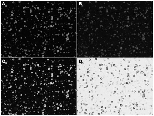
Embodiment 1
[0067] Example 1: Preparation of ultrasound contrast agent imitating leukocyte microbubbles
[0068] Rats were intraperitoneally injected with LPS 1.5 mg / kg. Six hours later, after sevoflurane inhalation anesthesia, the abdominal cavity was opened to expose the abdominal aorta to collect blood in anticoagulant blood collection tubes. Take another 50ml centrifuge tube and add the same volume of rat peripheral blood leukocyte separation medium (Solarbio) as the blood sample; use a Pasteur pipette to draw the blood sample, spread it carefully on the surface of the separation medium, and centrifuge at 1000g for 30min. After centrifugation, two ring-shaped milky white cell layers will appear in the centrifuge tube, the upper layer of cells is a mononuclear cell layer, and the lower layer of cells is a white blood cell layer. Use a pipette to carefully absorb the white blood cell layer in the separation solution, add 3 times the cell volume of red blood cell lysate (Solarbio), gentl...
Embodiment 2
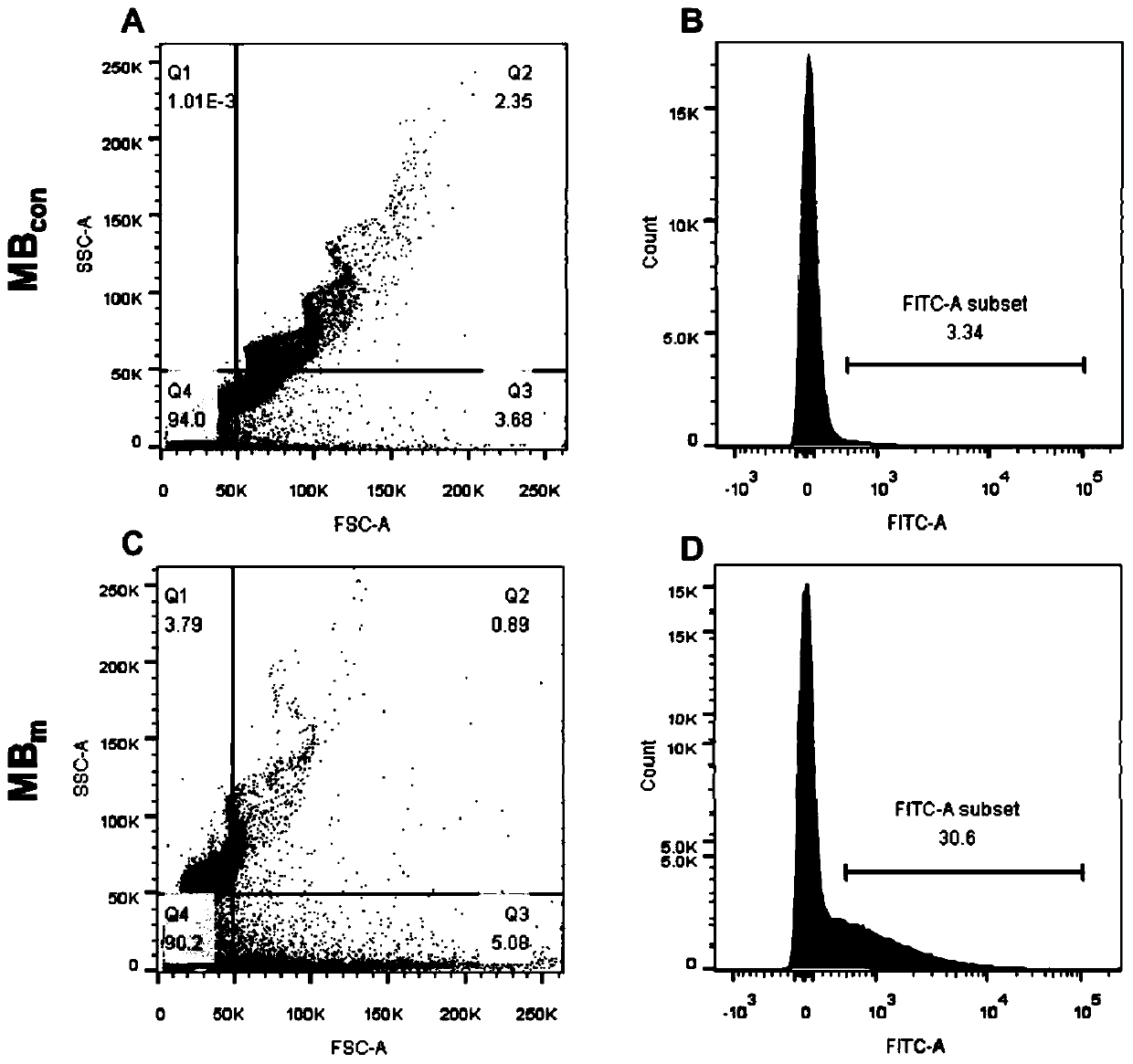
[0076] Example 2: Detection of Biomimetic Microvesicle Targeting Ligands
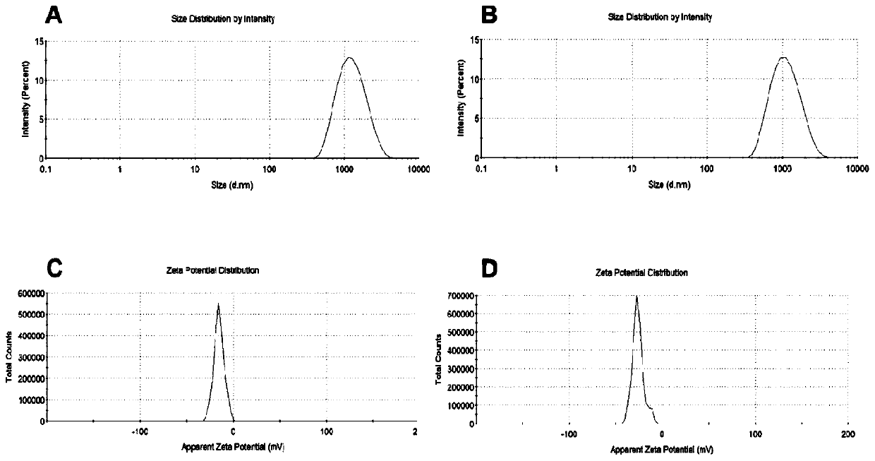
[0077] The control and biomimetic microbubbles prepared above were purified by centrifugation (2000 rpm, 2 min), the lower layer liquid was discarded, resuspended in 500 μl PBS, and 4.5 μg CD45 flow cytometry antibody was added, and incubated at room temperature for 30 minutes. Then centrifuge again (2000rpm, 2min), discard the lower liquid to remove free antibody, add 500μl PBS to resuspend, dilute to 10 5 cells / ml, the expression of microvesicle CD45 was detected by flow cytometry.
[0078] The results showed that: biomimetic microbubbles (MB m ) CD45 expression level (30.6%) was significantly higher than control microvesicles (MB con )(3.34%)( image 3 ), indicating that the biomimetic microvesicles carry the leukocyte membrane-specific protein CD45.
[0079] The control and biomimetic microbubbles prepared above were purified by centrifugation (2000 rpm, 2 min), the lower layer liquid was discar...
Embodiment 3
[0082] Example 3: Ultrasound imaging ability detection of bionic microbubble ultrasound contrast agent
[0083] Establish rat liver ischemia-reperfusion injury (ischemia-reperfusion injury, IRI) model: rats were fasted for 8-12 hours before surgery, and anesthetized by intraperitoneal injection of a compound solution of ketamine (60mg / kg) and xylazine (100mg / kg) . Fix the rat on the operating table, make a median abdominal incision, expose the abdominal cavity, dissociate the hilar blood vessels, clamp the blood flow in the left lobe and middle lobe of the liver with blood vessel clips, remove the blood vessel clips after 60 min, and suture the incision. The operation of ischemia 0min in the control group was performed after laparotomy.
[0084] After the animals were anesthetized, they were fixed on the operating table. Cut the suture along the midline abdominal incision, and fix the ultrasound probe (color Doppler ultrasound diagnostic system, Philips EPQ7, 5-12L high-freq...
PUM
 Abstract
Description
Claims
Application Information
 - R&D Engineer
- R&D Manager
- IP Professional
- Industry Leading Data Capabilities
- Powerful AI technology
- Patent DNA Extraction
Browse by: Latest US Patents, China's latest patents, Technical Efficacy Thesaurus, Application Domain, Technology Topic, Popular Technical Reports.
© 2024 PatSnap. All rights reserved.Legal|Privacy policy|Modern Slavery Act Transparency Statement|Sitemap|About US| Contact US: help@patsnap.com