Preparation method and application of rat chronic dorsal root neurothlipsis model
A technology of dorsal root nerve and model, applied in the field of pathology and zoology, can solve the problems of severe animal damage and difficult operation, and achieve the effect of small animal damage, good stability, and easy objective measurement.
- Summary
- Abstract
- Description
- Claims
- Application Information
AI Technical Summary
Problems solved by technology
Method used
Image


Examples
Embodiment 1
[0033] Example 1. Preparation of model
[0034] 1. Rats (220-250g) were anesthetized by intraperitoneal injection of 1% sodium pentobarbital (40mg / kg), the back was shaved, the back skin was disinfected with iodophor, fixed on the animal operating table in the prone position and covered with sterile towels.
[0035] 2. Make a 2-3cm longitudinal incision on the left side of the rat's lumbar 4 to sacral 1 level, cut the skin, and bluntly separate the paraspinal muscles to the top of the transverse process, exposing the lumbar 4 intervertebral foramen and lumbar 5 intervertebral foramen ( The paraspinal muscles can be kept separated by self-made hooks).
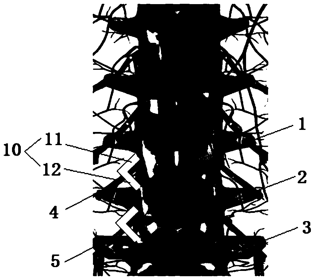
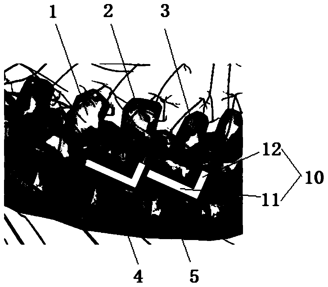
[0036] 3. Insert two L-shaped rods 10 with the first end 11 of 4mm, the second end 12 of 2mm and the diameter of 0.6mm respectively into the lumbar 4 intervertebral foramen and lumbar 5 intervertebral foramen at a certain angle. The two ends 12 are placed outside the lumbar 4 intervertebral foramen and lumbar 5 intervertebral foramen....
Embodiment 2
[0040] Example 2. Effect detection of the model
[0041] The present invention has verified the effect of the model through experiments. Through 28 days of observation, it was determined that the model obtained in Example 1 stably and accurately simulated the claudication and pain symptoms after the lumbar disc herniated and compressed the nerve root. image 3 It shows that the postoperative rat's hind paw on the operating side (left side) (see part A) did not grasp the net frame with the other three paws, but curled up, showing the performance of not daring to bear weight.
[0042] Table 1 Rat foot withdrawal threshold on different days after operation (unit: g)
[0043]
[0044] Table 1 shows the specific mean value and standard error of the withdrawal threshold of both lower limbs of 11 rats within 28 days after the operation. It can be seen that the withdrawal threshold of the hind paw on the surgical side was significantly lower than that before and on the contralateral hind paw...
Embodiment 3
[0046] Example 3: Application of the model
[0047] The two groups of rats were modeled using the method described in Example 1. The material of the L-shaped rods was H62 brass or polylactic acid, which is not interfered by magnetic fields, and will not cause chemical stimulation to nerves when placed in the body. A material with a certain degree of plasticity. On the seventh day after modeling, rats were treated with repeated transcranial magnetic stimulation (rTMS). One group received real magnetic stimulation, which was the magnetic stimulation group; the other group received fake magnetic stimulation, which was the fake stimulation group. The results showed that the withdrawal threshold of rats in the magnetic stimulation group was significantly higher than that in the sham stimulation group. Figure 5 Shown. This indicates that magnetic stimulation relieves the pain caused by nerve compression, and this model can be used for research on magnetic stimulation therapy.
PUM
| Property | Measurement | Unit |
|---|---|---|
| Diameter | aaaaa | aaaaa |
| Diameter | aaaaa | aaaaa |
| Diameter | aaaaa | aaaaa |
Abstract
Description
Claims
Application Information
 - R&D
- Intellectual Property
- Life Sciences
- Materials
- Tech Scout
- Unparalleled Data Quality
- Higher Quality Content
- 60% Fewer Hallucinations
Browse by: Latest US Patents, China's latest patents, Technical Efficacy Thesaurus, Application Domain, Technology Topic, Popular Technical Reports.
© 2025 PatSnap. All rights reserved.Legal|Privacy policy|Modern Slavery Act Transparency Statement|Sitemap|About US| Contact US: help@patsnap.com