
Method for separate-culturing cardiomyocytes

A cardiomyocyte, separation and culture technology, applied in the field of separation and culture of cardiomyocytes in vitro, can solve problems such as long time, unfavorable maintenance of cardiomyocyte activity, reduction of cardiomyocyte activity, etc., to enhance vitality, reduce cell damage, and avoid overdigestion. Effect

Pending Publication Date: 2020-05-15
GUANGDONG BOXI BIO TECH CO LTD
View PDF6 Cites 3 Cited by
  • Summary
  • Abstract
  • Description
  • Claims
  • Application Information

AI Technical Summary

Problems solved by technology

However, the activity of cardiomyocytes will gradually decrease with the extension of time in vitro
The compound enzyme does not contain various components of the culture medium necessary for the survival of cardiomyocytes, and the digestion time at 4°C is too long, which is not conducive to the maintenance of the activity of cardiomyocytes after separation

Method used


Image

  • Method for separate-culturing cardiomyocytes
  • Method for separate-culturing cardiomyocytes
  • Method for separate-culturing cardiomyocytes

Examples


Embodiment 1

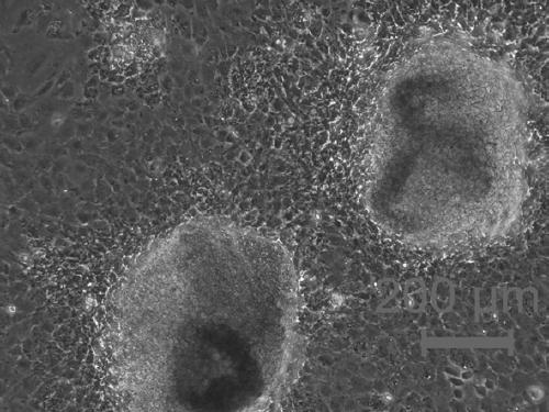
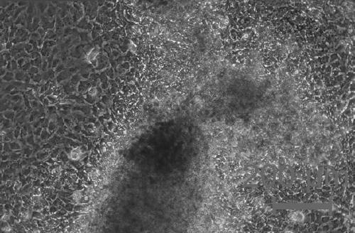
[0029] This embodiment provides a method for isolating and culturing cardiomyocytes of 6-month-old adult rats, which includes the following steps: placing the cardiomyocytes in PBS liquid for cleaning until the residual blood is cleaned; cut into 0.5mm 3 Add 3mL of type Ⅰ collagenase, shake and digest at 37°C for 2 hours; add 10 times the volume of type Ⅰ collagenase in normal temperature PBS liquid to dilute and stop after digestion; centrifuge at 1200rpm / min for 5min, discard the supernatant . The cell pellet was resuspended with 3 mL of trypsin, and the elbow pipette was pipetted 50 times. After the digestion solution became turbid, the trypsin digestion reaction was terminated with culture medium, centrifuged at 1200 rpm / min for 5 minutes, and the supernatant was discarded; the cell pellet was resuspended with culture medium. cells according to 4×10 5 cells / cm 2 Inoculate cell culture flasks, cell culture dishes or multi-well plates for cell culture at 37°C, 5% CO 2 cu...

Embodiment 2

[0031] This embodiment provides a method for isolating and culturing cardiomyocytes of New Zealand baby rabbits within 3 days after birth, which includes the following steps: placing the cardiomyocytes in PBS liquid for cleaning until the residual blood is cleaned, and then using ophthalmic scissors to clean the cardiomyocytes Heart tissue cut into 0.5mm 3 Add 2mL of type Ⅰ collagenase, shake and digest at 37°C for 1.5 hours, add 10 times the volume of type Ⅰ collagenase in normal temperature PBS liquid to dilute and stop after digestion, centrifuge at 1200rpm / min for 5min, discard the supernatant ;The cell pellet was resuspended with 2mL of trypsin, and pipetted 10 times with an elbow pipette. After the digestion solution became turbid, the trypsin digestion reaction was terminated with culture medium, centrifuged at 1200rpm / min for 5min, and the supernatant was discarded. The cell pellet was resuspended with culture medium, and the cells were divided into 2×10 5 cells / cm 2...


PUM


Abstract

The invention provides a method for separate-culturing cardiomyocytes. The method includes the following steps that (1) heart tissue is taken, and PBS liquid is used to wash away residual blood; the heart tissue is cut into tissue blocks of 0.1-1mm<3> with ophthalmic scissors, 0.5-5ml of collagenase I is added, and shaking digesting is carried out at 36-37.5 DEG C for 0.5-3 hours; after digestion,2.5-50mL of PBS is added for dilution and stop, centrifuging is carried out at 1200rpm / min for 5 min, and a supernatant is discarded to obtain a cell pellet; (2) the cell pellet is resuspended with 0.5-5mL pancreatin, an elbow pipette is used for blowing-beating for 10-300 times, after digestion fluid is turbid, a culture medium is added to stop pancreatin digestion, and centrifuging is carried out at 1200rpm / min for 5 min, and a supernatant is discarded to obtain primary cardiomyocytes; and (3) after the primary cardiomyocytes is resuspended in the culture medium, the primary cardiomyocytesare inoculated into the culture medium according to 0.1-10x10<5> cells / cm<2>, and the obtained mixture is placed in a culture incubator of 37 DEG C and 5%CO2 for culturing to obtain the cardiomyocytes.

Description

technical field [0001] The invention belongs to the technical field of cell culture, and in particular relates to a method for separating and culturing cardiomyocytes in vitro. Background technique [0002] Cardiomyocytes are terminally differentiated mature cells that play an important role in maintaining the normal shape and structure of the heart. In recent years, cardiomyocytes cultured in vitro have been mostly used as research objects in the study of the physiological and pathological development of the heart, as well as in the development and pharmacological exploration of cardiovascular-related drugs. [0003] The existing methods are mainly aimed at the isolation of cardiomyocytes from fetal rats or neonatal mice within 3 days of birth, and are not suitable for the isolation of cardiomyocytes from adult rats, mice, and more animal sources. However, at the cell and molecular level of cardiomyocytes, it is often necessary to obtain cardiomyocytes from adult animals o...

Claims


Application Information

Patent Timeline
Patent Type & Authority Applications(China)
IPC IPC(8): C12N5/077
CPCC12N5/0657C12N2509/00
Inventor 许珊珊张勇杰卢永波
Owner GUANGDONG BOXI BIO TECH CO LTD
Learn More