Transcranial electrical stimulating assembly, device, system and application
An electrical stimulation and transcranial technology, applied in applications, electrodes, electrotherapy, etc., can solve the problems of lack of transcranial electrical stimulation devices, achieve the effects of reducing pulsation, realizing long-term living imaging, and preventing external environmental infection
- Summary
- Abstract
- Description
- Claims
- Application Information
AI Technical Summary
Problems solved by technology
Method used
Image
Examples
Embodiment 1
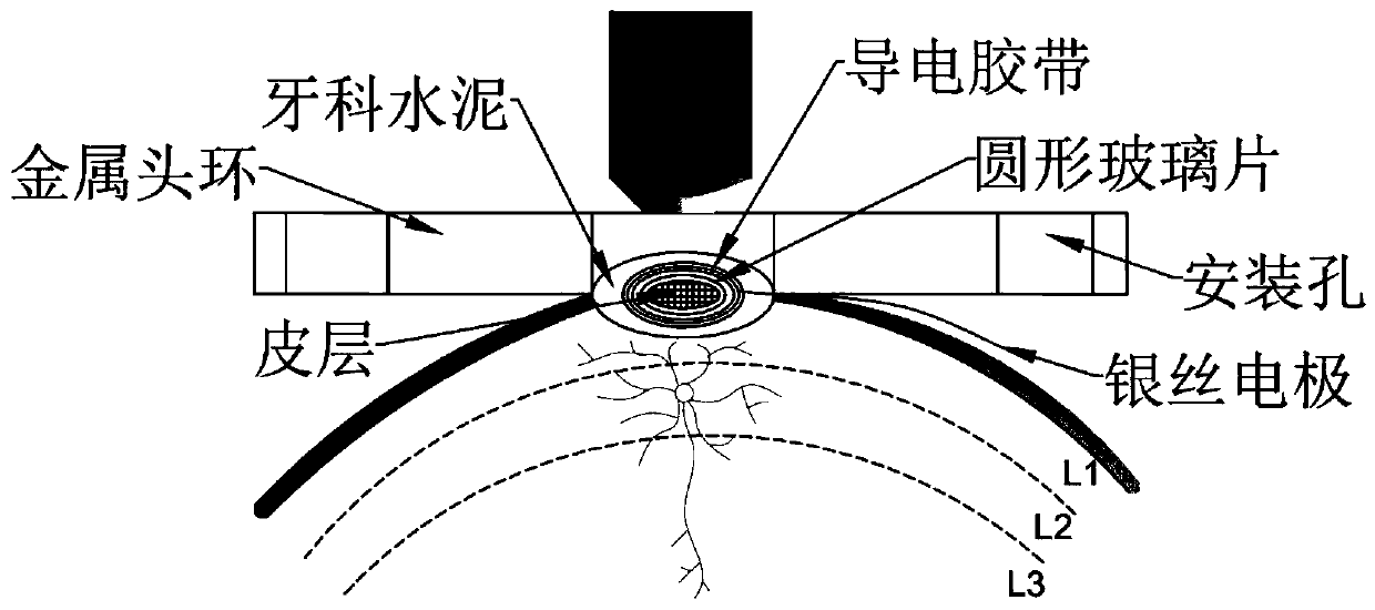
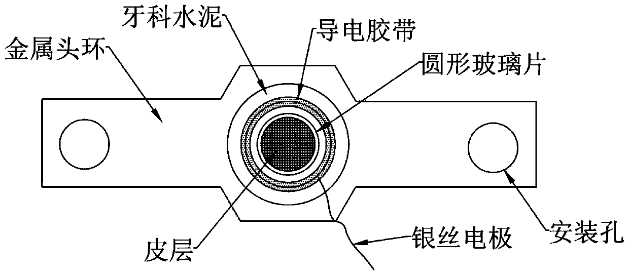
[0041] This embodiment provides a transcranial electrical stimulation component. refer to figure 1 and figure 2 As shown, there are imaging holes and installation holes on the metal head ring. The round glass piece covers the animal craniotomy cortex. There are silver wires and conductive tape around the glass piece. The round glass piece is covered with Compaite medical glue and dental cement fixed on the animal skull.
Embodiment 2
[0043] This embodiment provides the method for installing and using the transcranial electrical stimulation device in Embodiment 1, which specifically includes the following steps:
[0044] (1) Surgical instruments and transcranial electrical stimulation devices are sterilized in an autoclave;
[0045] (2) 8-week-old mice were fed normally, anesthetized with isoflurane gas, and fixed on a stereotaxic apparatus. Remove the local scalp and fascia, rinse with low-concentration cephalosporin (cephalosporin and saline ratio 1:250), and determine the craniotomy area of the cranial window, the diameter of which can be 3-4mm. After craniotomy, the virus vector AAV-Syn-GCaMP6f-WPRE was injected into the exposed cortex to transfect the cortical neurons, thereby expressing the GCamp6f fluorescent protein in the neurons, and then realizing the later two-photon calcium imaging to study the calcium activity of the neurons Variety. After viral vector injection, the round glass slide and ...
experiment example 1
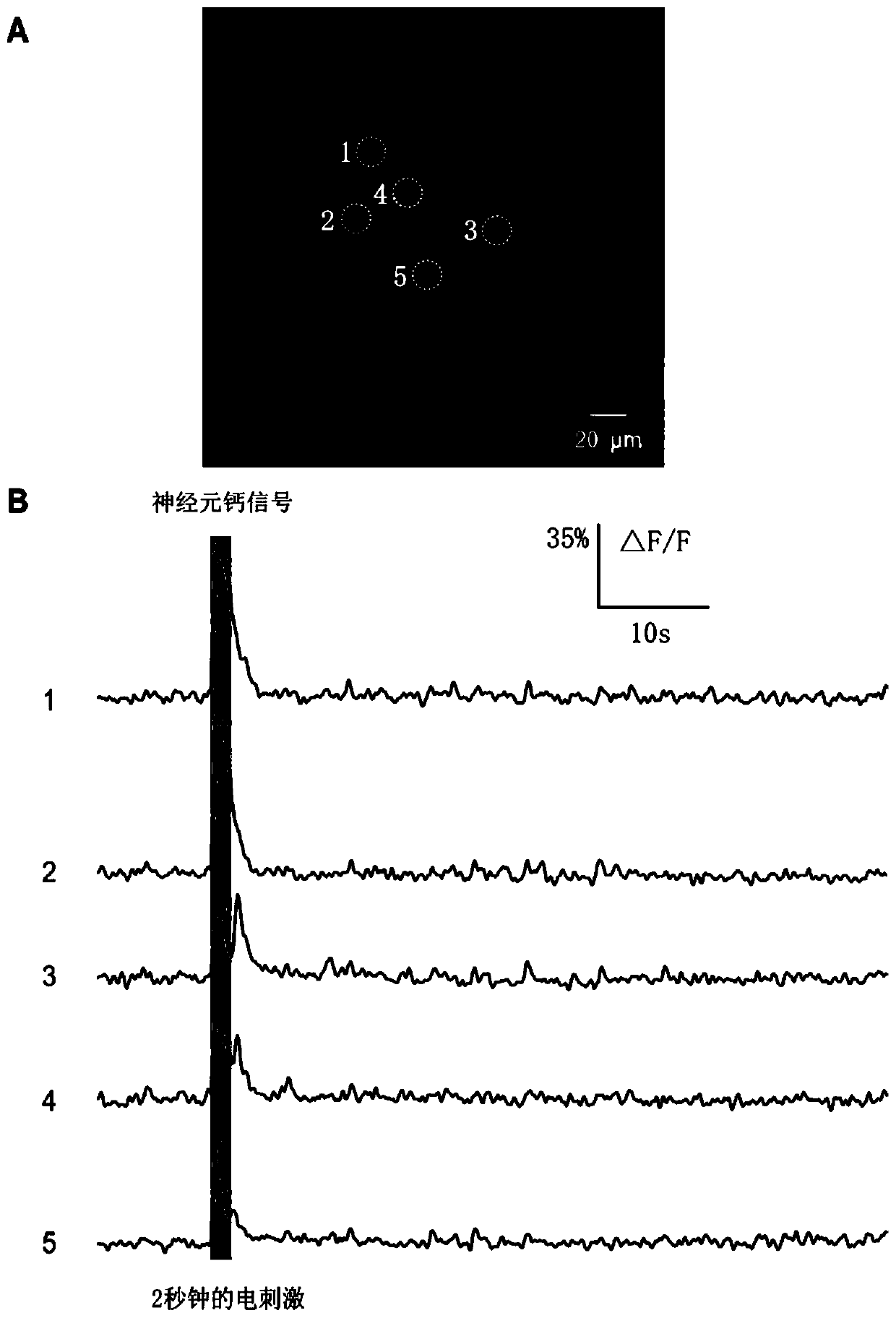
[0051] This experimental example is an electrical stimulation and two-photon imaging experiment performed using the mouse model in Example 2. The expression effect of GCamp6f fluorescent protein in neurons after 3 weeks of mouse cortical injection image 3 As shown in Figure A, it can be seen from the figure that neurons normally express GCamp6f fluorescent protein. Set the electrical stimulation parameters as bidirectional pulses, with a frequency of 50 Hz, a current intensity of 0.3 mA, and a duration of 2 seconds. Experimental results refer to image 3 As shown in panel B in , the calcium signal of neurons evoked by electrical stimulation through the silver wire changed significantly.
PUM
| Property | Measurement | Unit |
|---|---|---|
| Diameter | aaaaa | aaaaa |
Abstract
Description
Claims
Application Information
 - R&D
- Intellectual Property
- Life Sciences
- Materials
- Tech Scout
- Unparalleled Data Quality
- Higher Quality Content
- 60% Fewer Hallucinations
Browse by: Latest US Patents, China's latest patents, Technical Efficacy Thesaurus, Application Domain, Technology Topic, Popular Technical Reports.
© 2025 PatSnap. All rights reserved.Legal|Privacy policy|Modern Slavery Act Transparency Statement|Sitemap|About US| Contact US: help@patsnap.com


