Detection method for medical image segmentation and medical image segmentation method and device
A technology of medical images and detection methods, applied in the field of image processing, can solve the problem of not forming end-to-end
- Summary
- Abstract
- Description
- Claims
- Application Information
AI Technical Summary
Problems solved by technology
Method used
Image

Examples
Embodiment 1
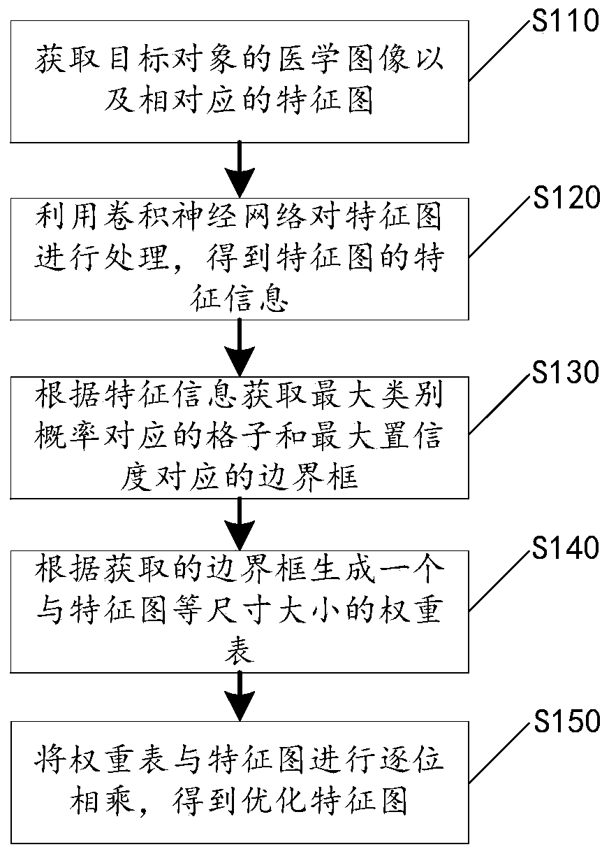
[0036] Please refer to figure 1 , the present application discloses a detection method for medical image segmentation, which includes steps S110-S150, which will be described respectively below.
[0037] Step S110, acquiring a medical image of a target object and a feature map corresponding to the medical image, where the target object is a lesion formed on a tissue or organ.
[0038] In this embodiment, common medical imaging equipment can be used to shoot specific tissues and organs of the patient, so as to obtain medical images such as CT images, MRI images, PET images, and DSA images, and the target object can be kidney tumors , Lung tumors, liver tumors, stomach tumors and other types of lesions, these lesions are often formed on the internal tissues and organs of patients, and have the characteristics of small targets (often in millimeters in diameter) and low discrimination.
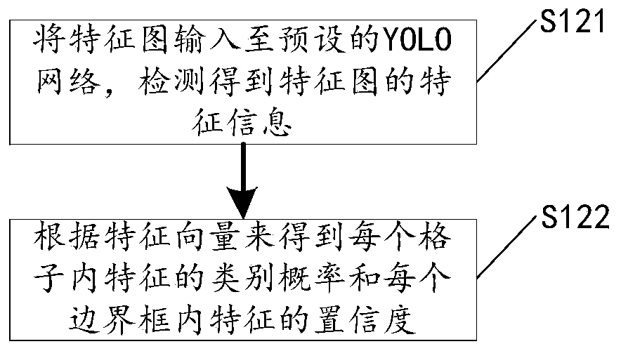
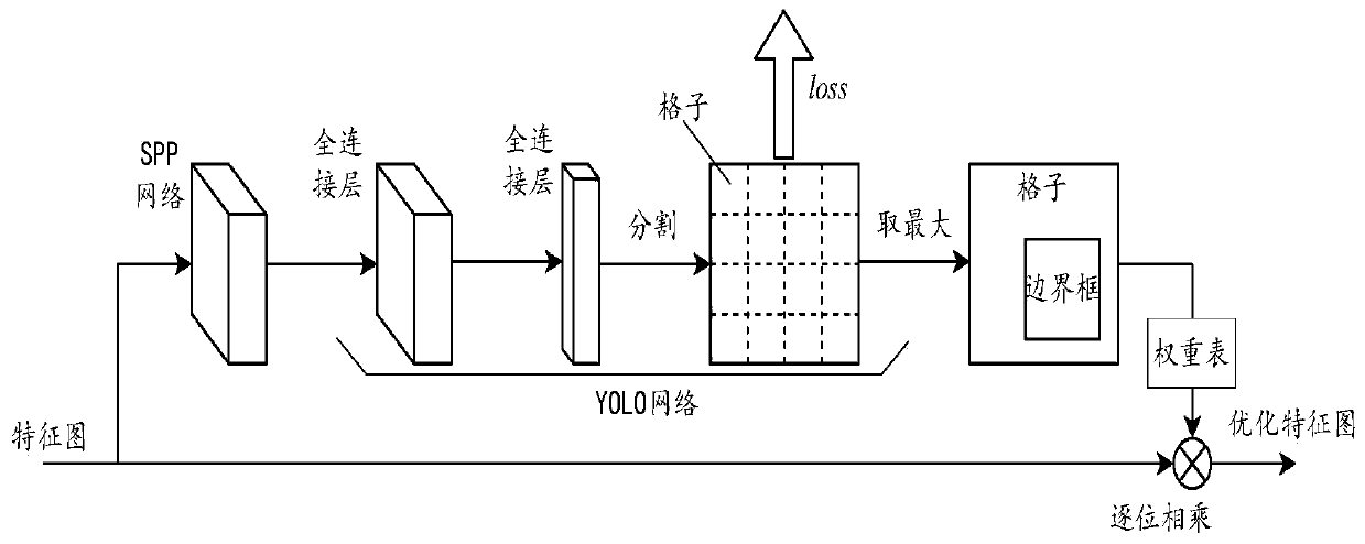
[0039] Step S120, using the convolutional neural network to process the feature map to obtain...
Embodiment 2
[0057] Please refer to Figure 4 , on the basis of the detection method for medical image segmentation disclosed in Embodiment 1, a medical image segmentation method is also disclosed, the medical image segmentation method includes steps S210-S230, which will be described respectively below.
[0058] Step S210, acquiring a medical image of a target object, where the target object is a lesion formed on a tissue or organ.
[0059] For example, common medical imaging equipment can be used to shoot specific tissues and organs of patients to obtain medical images such as CT images, MRI images, PET images, and DSA images, and the target objects can be kidney tumors, lung tumors, Liver tumors, gastric tumors and other types of lesions, which are often formed on the internal tissues and organs of patients, have the characteristics of small targets (often in millimeters in diameter) and low discrimination.
[0060] In step S220, the medical image is input into a pre-established lesion...
Embodiment 3
[0090] Please refer to Figure 8 , on the basis of the medical image segmentation method for medical images disclosed in Embodiment 2, this application discloses a target recognition device 4 for medical images, which mainly includes an acquisition unit 41, a model processing unit 42, a recognition Unit 43 will be described separately below.
[0091] The acquiring unit 41 is configured to acquire a medical image of a target object, where the target object is a lesion formed on a tissue or organ. In this embodiment, common medical imaging equipment can be used to shoot specific tissues and organs of the patient, so as to obtain medical images such as CT images, MRI images, PET images, and DSA images, and the target object can be kidney tumors , Lung tumors, liver tumors, stomach tumors and other types of lesions, these lesions are often formed on the internal tissues and organs of patients, and have the characteristics of small targets (often in millimeters in diameter) and lo...
PUM
 Abstract
Description
Claims
Application Information
 - R&D
- Intellectual Property
- Life Sciences
- Materials
- Tech Scout
- Unparalleled Data Quality
- Higher Quality Content
- 60% Fewer Hallucinations
Browse by: Latest US Patents, China's latest patents, Technical Efficacy Thesaurus, Application Domain, Technology Topic, Popular Technical Reports.
© 2025 PatSnap. All rights reserved.Legal|Privacy policy|Modern Slavery Act Transparency Statement|Sitemap|About US| Contact US: help@patsnap.com