Hip joint imaging method and hip joint imaging system

An imaging system and imaging method technology, applied in the field of medical devices, can solve the problems of deviation in measurement and high requirements for slice images

Pending Publication Date: 2019-12-24
PEKING UNIV THIRD HOSPITAL +2
View PDF6 Cites 8 Cited by
  • Summary
  • Abstract
  • Description
  • Claims
  • Application Information

AI Technical Summary

Problems solved by technology

[0005] Therefore, when performing DDH ultrasonography according to the Graf method, the requirements for section images are high, and if the sections are not standard, there will be deviations in the measurement

Method used


Image

  • Hip joint imaging method and hip joint imaging system
  • Hip joint imaging method and hip joint imaging system
  • Hip joint imaging method and hip joint imaging system

Examples


Embodiment Construction

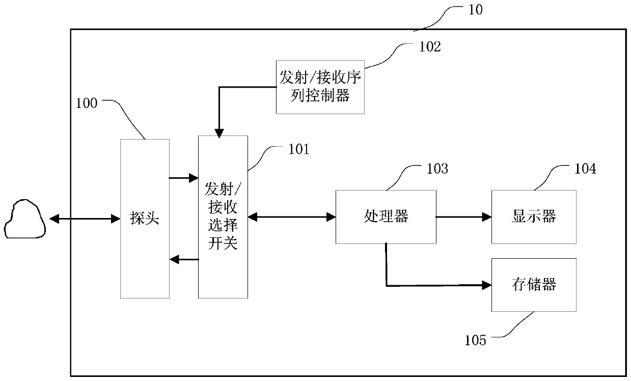
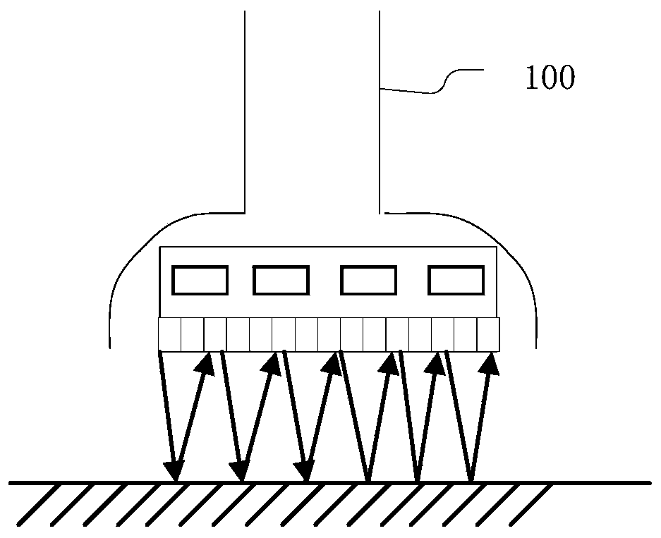
[0030] The present application provides a hip joint imaging method and a hip joint imaging system, which are used to obtain a standard section view of the hip joint.

[0031] The terms "first", "second", "third", "fourth", etc. (if any) in the specification and claims of the present application and the above drawings are used to distinguish similar objects, and not necessarily Used to describe a specific sequence or sequence. It is to be understood that the terms so used are interchangeable under appropriate circumstances such that the embodiments described herein can be practiced in sequences other than those illustrated or described herein. Furthermore, the terms "comprising" and "having", as well as any variations thereof, are intended to cover a non-exclusive inclusion, for example, a process, method, system, product or device comprising a sequence of steps or elements is not necessarily limited to the expressly listed instead, may include other steps or elements not expl...


PUM


Abstract

The invention provides a hip joint imaging method and a hip joint imaging system, which are used for obtaining a standard section diagram of a hip joint. The method comprises the following steps: acquiring volume data of the target hip joint; obtaining at least one section diagram of the target hip joint according to the volume data; analyzing the at least one section diagram to obtain analysis data of the at least one section diagram, wherein the analysis data is used for measuring a standard degree of the at least one section diagram; and determining at least one standard section diagram from the at least one section diagram according to the analysis data.

Description

technical field [0001] The present application relates to the field of medical devices, in particular to a hip joint imaging method and a hip joint imaging system. Background technique [0002] Ultrasound imaging is widely used in the analysis of various diseases. Ultrasound imaging can be performed on the hip joint to analyze the characteristics of the hip joint tissue and determine the health status of the hip joint tissue. [0003] For example, developmental dysplasia of the hip (DDH) is the most common hip disease in children. It is a general term for a series of hip abnormalities that exist at birth or continue to develop after birth: Includes stable hip with acetabular dysplasia, hip subluxation, complete hip dislocation that can be reduced, and complete dislocation that cannot be reduced. The currently recognized DDH treatment principle is early detection and early treatment. The earlier the treatment, the simpler the treatment method and the better the treatment ef...

Claims


Application Information

Patent Timeline
IPC IPC(8): A61B8/00A61B8/08
CPCA61B8/4411A61B8/5223A61B8/4444A61B8/461A61B8/0875
Inventor 崔立刚陈涛王勃丛龙飞朱磊
Owner PEKING UNIV THIRD HOSPITAL
Why Eureka
Try Eureka