Schistosoma japonicum katsurada antibody detection kit with detection protein having red fluorescence activity
A red fluorescent protein and antibody detection technology, applied in the field of immunological detection, can solve the problems of sensitivity improvement, achieve convenient storage and transportation, and have industrial effects
- Summary
- Abstract
- Description
- Claims
- Application Information
AI Technical Summary
Problems solved by technology
Method used
Image
Examples
Embodiment 1
[0035] The construction of embodiment 1 recombinant protein G
[0036] Escherichia coli BL21 was purchased from Nanjing Novizan Biotechnology Co., Ltd.; the plasmid pET-28a(+) was stored in the laboratory, and the bacterial liquid containing the C3 fragment[1] and the bacterial liquid containing the RFP sequence[2] were used for the experiment Room preservation.
[0037][1] Xu Rui, Zhao Dengyun, Hong Yang, Lu Ke, Li Hao, Lin Jiaojiao, Feng Jintao, Xu Yumei, Zhu Chuangang. Domain remodeling, expression and identification of streptococcal protein G [J]. Chinese Journal of Animal Infectious Diseases ,2015,23(05):46-52.
[0038] [2] Kang Zeran. Cloning, prokaryotic expression characteristics and biological information analysis of monomeric red fluorescent protein gene DsRed2[D]. Yanbian University, 2016.
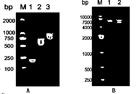
[0039] The fusion protein C3-RFP expressing recombinant G protein and red fluorescent protein was obtained by means of PCR, the nucleotide sequence of the fusion protein C3-RF...
Embodiment 2
[0041] Expression and purification of embodiment 2 recombinant protein G
[0042] 2.1 Expression of recombinant plasmids
[0043] phase
[0044] (1) Transfer the pET-28a(+)-C3-RFP recombinant plasmids with correct identification results into BL21(DE3), inoculate them in 5ml LB liquid medium containing Kan+, and place them in a shaking incubator at 37°C. Shake culture at 250rpm.
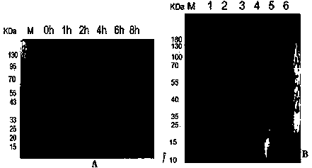
[0045] (2) When growing to the logarithmic phase (OD600 is about 0.6), add IPTG with a final concentration of 1 mmol / L to induce expression. Take 0.5ml bacterial liquid before induction and 1h, 2h, 4h, 6h, 8h after induction, and analyze the best induction time by SDS-PAGE electrophoresis. ( image 3 A)
[0046] Massive expression:
[0047] (1) Transform the pET-28a(+)-C3-RFP recombinant plasmids with correct identification results into BL21(DE3), inoculate them in 150ml LB liquid medium containing Kan+, and place them in a shaking incubator at 37°C. Shake culture at 250rpm.
[0048] (2) When ...
Embodiment 3
[0060] Example 3 Activity identification of C3-RFP recombinant protein
[0061] 3.1 Observation of fluorescence activity
[0062] Centrifuge the bacterial solution collected in the above phase at 10,000rpm for 5min, discard the supernatant, and add 200ul ddH 2 O resuspend the bacteria, after resuspension, pipette 10ul onto a clean glass slide, cover with a cover glass, excite with RFP excitation light under a fluorescent electron microscope, and observe whether the bacteria have red fluorescence. Fluorescence electron microscopy observations showed that ( Figure 4 ), the fluorescence of recombinant plasmid pET-28a(+)-C3-RFP successfully induced and expressed in E. coli BL21(DE3) increased with time at 1-8h, and the fluorescence reached the highest after 8h of induction and tended to be stable.
[0063] 3.2 Western blotting to detect the binding activity of recombinant protein and IgG
[0064] (1) Perform SDS-PAGE electrophoresis on the purified protein, then transfer the p...
PUM
 Abstract
Description
Claims
Application Information
 - Generate Ideas
- Intellectual Property
- Life Sciences
- Materials
- Tech Scout
- Unparalleled Data Quality
- Higher Quality Content
- 60% Fewer Hallucinations
Browse by: Latest US Patents, China's latest patents, Technical Efficacy Thesaurus, Application Domain, Technology Topic, Popular Technical Reports.
© 2025 PatSnap. All rights reserved.Legal|Privacy policy|Modern Slavery Act Transparency Statement|Sitemap|About US| Contact US: help@patsnap.com


