
A method for constructing sers spectral probe to detect breast cancer marker EGFR phosphorylated tyrosine

A breast cancer and phosphorylation technology, which is applied in measuring devices, biological tests, material inspection products, etc., can solve the problems of patients' risks and high prices, and achieve the effects of improving detection sensitivity, improving linear relationship, and ensuring reliability

Active Publication Date: 2022-05-03
FUJIAN NORMAL UNIV
View PDF5 Cites 0 Cited by
  • Summary
  • Abstract
  • Description
  • Claims
  • Application Information

AI Technical Summary

Problems solved by technology

However, it is invasive, risky to the patient and relatively expensive

Method used


Image

  • A method for constructing sers spectral probe to detect breast cancer marker EGFR phosphorylated tyrosine
  • A method for constructing sers spectral probe to detect breast cancer marker EGFR phosphorylated tyrosine
  • A method for constructing sers spectral probe to detect breast cancer marker EGFR phosphorylated tyrosine

Examples


Embodiment Construction

[0031] The present invention will be further described below in conjunction with specific embodiments and accompanying drawings.

[0032] 1. Reagents Chlorauric acid solution (HAuCl4), sodium citrate (Na3C6H5O7), p-mercaptobenzoic acid (4-MBA), p-mercaptobenzonitrile (4-MB), ascorbic acid, silver nitrate (AgNO3), phosphate buffer solution (PBS, PH=7), phosphorylated serine dipeptide (PS-S), phosphorylated threonine dipeptide (PT-T), single-stranded DNA (S1: AAAATGTCCAAGGCT-SH, S2: TTTTACAGGTTCCGA-SH), Bovine serum albumin (BSA), anti-EGFR phosphorylated tyrosine antibody (Anti-EGFR(Phospho-Tyr1172) antibody), antigen EGFR phosphorylated tyrosine polypeptide (DNPDY(P)QQDFFP), EGFR non-phosphotyrosine acid polypeptide (DNPDYQQDFFP). 2. Preparation of the solution

[0033] The preparation of 1% chloroauric acid solution, the specific steps are as follows: First, dissolve the chloroauric acid solid powder in a beaker with ultrapure water, then pour it into a 100mL volumetric fla...


PUM

PropertyMeasurementUnit
particle diameteraaaaaaaaaa
concentrationaaaaaaaaaa

Abstract

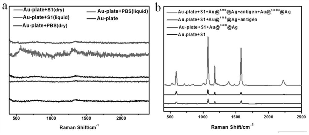
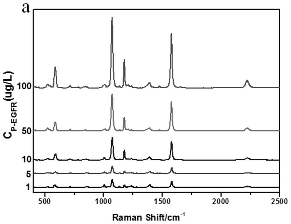
The invention discloses a method for constructing a SERS spectral probe to detect breast cancer marker EGFR phosphorylated tyrosine: a gold plate is used as a substrate, and core-shell structure nanoparticles are used as a substrate for SERS detection to develop high selectivity and high sensitivity A "sandwich" configuration probe molecule to detect the phosphorylation of breast cancer marker EGFR tyrosine. Using the 4‑MB Raman spectrum, the peak position in the Raman silent zone of biomolecules is 2221cm ‑1 The intensity of the SERS spectrum of the marker molecule was used as an internal standard to normalize the intensity of the spectral signal of the marker molecule 4-MBA, which can avoid the influence of the Raman signal in the biomolecular fingerprint region on the quantitative detection, and make the concentration of the EGFR phosphorylated tyrosine solution and the SERS intensity The linear relationship is greatly improved, further improving the detection sensitivity of the SERS spectral probe. 4-MB and 4-MBA are located in the middle layer between the gold core and the silver shell, which ensures that its properties, spatial structure and quantity remain unchanged, thereby ensuring the reliability of the data. The method of the present invention shows great potential in ultrasensitive detection of cancer markers.

Description

technical field [0001] The invention belongs to the field of phosphorylation detection of biomedical cancer markers, and in particular relates to a method for constructing a SERS spectral probe to detect phosphorylated tyrosine of breast cancer marker EGFR. Background technique [0002] Breast cancer is the second most common cancer in the world, accounting for 25% of all cancers, and breast cancer is also the most common malignant tumor in women in the world. Women over 45 years old. Epidermal growth factor receptor (EGFR) is one of the specific protein biomarkers of breast cancer. It is a transmembrane glycoprotein located on the cell surface. It is a complex with tyrosine kinase and epidermal growth factor and transforming growth factor (TGFa). Upon binding, activates and induces cell proliferation and differentiation. EGFR is overexpressed in many epithelial tumors (>75.3µg·L -1 ), such as breast cancer, gastric cancer, esophageal cancer, prostate cancer, etc. [...

Claims


Application Information

Patent Timeline
Patent Type & Authority Patents(China)
IPC IPC(8): G01N33/68G01N33/58G01N33/574G01N21/65
CPCG01N33/6872G01N33/57415G01N33/57484G01N33/58G01N21/658
Inventor 冯尚源刘秀杰卢玉栋
Owner FUJIAN NORMAL UNIV
Learn More