
Auxiliary analysis method and system for pathological image of thyroid cancer cells based on deep learning

A cytopathology and deep learning technology, applied in medical images, computer-aided medical procedures, informatics, etc., can solve problems such as inability to apply cytopathology and histopathology, and inability to assist full-section digital pathology analysis, achieving convenient management and improving Efficiency, the effect of improving the resolving power

Inactive Publication Date: 2019-10-15
台州市中心医院
View PDF6 Cites 9 Cited by
  • Summary
  • Abstract
  • Description
  • Claims
  • Application Information

AI Technical Summary

Problems solved by technology

However, the above scheme can only improve the efficiency and accuracy of image recognition, and cannot assist the analysis of full-slice digital pathology with a very large amount of data, and cannot be applied to full-slice image analysis of cytopathology and histopathology due to the large amount of data.

Method used


Image

  • Auxiliary analysis method and system for pathological image of thyroid cancer cells based on deep learning
  • Auxiliary analysis method and system for pathological image of thyroid cancer cells based on deep learning
  • Auxiliary analysis method and system for pathological image of thyroid cancer cells based on deep learning

Examples


Embodiment 1

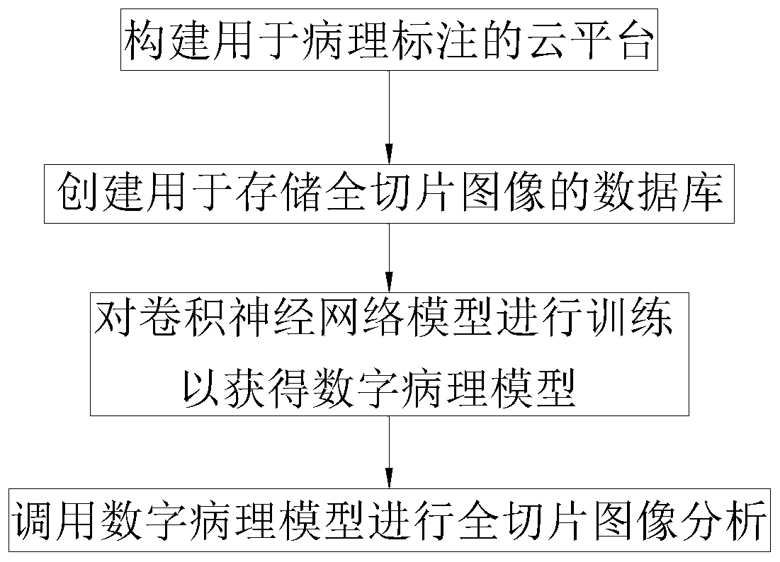
[0058] Such as figure 1 As shown, this embodiment discloses a method for auxiliary analysis of pathological images of thyroid cancer cells based on deep learning, mainly for the auxiliary analysis of pathological images of papillary thyroid cancer cells, specifically including the following steps:

[0059] S1. Build a cloud platform for digital pathology annotation to obtain annotated training set, verification set and test set;

[0060] S2. Create a database for storing pathological digital full slice images;

[0061] S3. Using the training set, verification set and test set to train the convolutional neural network model to obtain a digital pathology model;

[0062] S4. The digital pathological model is invoked by the decision-making system to obtain the digital pathological image analysis result of the pathological digital full slice image.

[0063] Different from traditional images, full slice data is very large. Generally, the size of ordinary images is on the order of ...

Embodiment 2

[0097] Such as Figure 10 As shown, this embodiment discloses a pathological image auxiliary analysis system based on the deep learning-based auxiliary analysis method for pathological maps of thyroid cancer cells described in Embodiment 1, including a cloud platform 1, a database 2, a deep learning model 3 and a decision-making System 4, where,

[0098] Cloud platform 1 is used to receive pathological digital full slice images from the database, and for professional doctors to digitally label the pathological digital full slice images in the platform to obtain marked training samples, namely training set, verification set and test set set;

[0099] Database 2 is used to receive and store pathological digital full slice images and marked training samples, the training samples include a training set, a verification set and a test set;

[0100] Deep learning model 3, for adopting training set, verification set and test set to carry out model training, verification and test to ...


PUM


Abstract

The invention provides an auxiliary analysis method and system for a pathological image of thyroid cancer cells based on deep learning. The method comprises the following steps: S1, constructing a cloud platform for digital pathological labeling to obtain a labeled training set, a labeled verification set and a labeled test set; S2, creating a database for storing a pathological digital full-sliceimage; S3, training a convolutional neural network model by using the training set, the verification set and the test set to obtain a digital pathological model; S4, calling a digital pathological model to carry out digital pathological image analysis on the pathological digital full-slice image. The invention provides the cloud platform for data labeling, the data transmission and labeling are carried out on line, and there is no special equipment requirement, thereby improving the labeling and data transmission efficiency; the database used for storing the full-slice image is constructed, the corresponding pathological digital full-slice images are obtained from the database in the labeling process, the training process and the subsequent analysis and detection process, thereby achieving the full-slice digital pathology analysis with a very large auxiliary data flow.

Description

technical field [0001] The invention belongs to the technical field of auxiliary analysis of pathological graphs, and in particular relates to a method and system for auxiliary analysis of pathological graphs of thyroid cancer cells based on deep learning. Background technique [0002] The final diagnosis of thyroid nodules and other lesions requires pathological diagnosis. Fine-needle aspiration cytology (FNA) is economical, easy to operate, less invasive, and has a high accuracy rate, which can improve the accuracy of lesion diagnosis. However, due to the small amount of tissue and its cellular components in fine-needle aspiration, most of the tissue morphology and intercellular matrix structure in the specimen are lost, and it is related to the experience of puncture and cytological diagnosis. Too small nodules and too thin needles may It will lead to insufficient or inaccurate sampling of the lesion. Due to the small amount of tissue taken, the difficulty in making and ...

Claims


Application Information

Patent Timeline
IPC IPC(8): G16H30/20G16H50/20G06K9/62G06F16/58G06F16/51G06N3/04
CPCG16H30/20G16H50/20G06F16/5866G06F16/51G06N3/045G06F18/241G06F18/214
Inventor 卢洪胜陈琪戴岳楚
Owner 台州市中心医院
Learn More