
Detection method for visual recognition of microRNA (Ribonucleic Acid) on the basis of G-quadruplex probe structure unwinding

A quadruplex and probe technology, applied in biochemical equipment and methods, microbial determination/inspection, etc., can solve the problems of high production cost, low abundance, and can not fully meet application requirements, and achieve the effect of simple design

Inactive Publication Date: 2019-09-13
INST OF CHEM CHINESE ACAD OF SCI
View PDF3 Cites 2 Cited by
  • Summary
  • Abstract
  • Description
  • Claims
  • Application Information

AI Technical Summary

Problems solved by technology

Since miRNA itself has the characteristics of short length, low abundance, easy degradation, and high similarity to homologous miRNAs, most studies are now devoted to signal amplification or target conversion methods to compensate for the defects of miRNAs, including polymerase chain reaction ( PCR), rolling circle amplification reaction (RCA), nanoparticle aggregation and fluorescence energy transfer, etc., but these methods have complicated reaction processes and high production costs, and cannot fully meet the application requirements.

Method used


Image

  • Detection method for visual recognition of microRNA (Ribonucleic Acid) on the basis of G-quadruplex probe structure unwinding
  • Detection method for visual recognition of microRNA (Ribonucleic Acid) on the basis of G-quadruplex probe structure unwinding
  • Detection method for visual recognition of microRNA (Ribonucleic Acid) on the basis of G-quadruplex probe structure unwinding

Examples


Embodiment 1

[0051] Embodiment 1, the design of probe and the establishment of detection method

[0052] The G-quadruplex probe designed by the present invention includes a binding segment and a signal segment, and the binding segment is a DNA fragment that can specifically recognize and bind to the target miRNA (the binding segment is reversely complementary to the full-length sequence of the target miRNA) , the signal segment is a DNA segment that can form a G-quadruplex structure for outputting a signal.

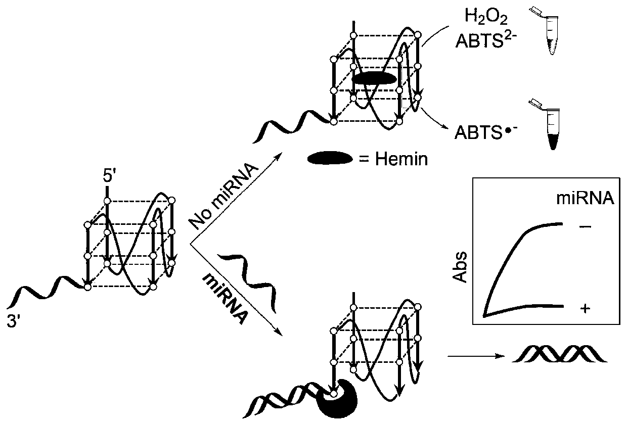
[0053] The detection principle of the present invention sees figure 1 . When there is no target miRNA molecule, the G-quadruplex structure in the probe can combine with heme to form DNAzyme, which can catalyze H 2 o 2 Oxidation of ABTS [2,2′-hydrazine-bis(3-ethylbenzothiazoline-6-sulfonic acid) diamine salt] reaction, the color of the solution changes from colorless to obvious green, which is characteristic in the absorption spectrum The absorption peak is 420nm; in the presence of ...

Embodiment 2

[0073] Example 2, G-quadruplex probe design using miRNA21 as target molecule

[0074] The G-quadruplex probe MG41 was designed with miRNA21 as the target molecule, as shown in Table 1.

[0075] Table 1 The sequence of G-quadruplex probe and miR21

[0076]

[0077] In the probe MG41, the 1st to 19th positions from the 5' end are the signal segment, and the 20th to 41st positions are the binding segment.

Embodiment 3

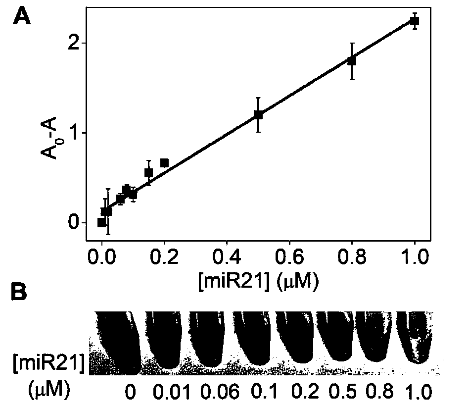
[0078] Embodiment 3, sensitivity

[0079] 1. Heat the MG41 probe prepared in Example 1 at 95°C for 5 minutes, and then place it on ice to anneal and cool down.

[0080] 2. Configure the reaction system as follows, and perform the hybridization reaction at 37°C for 80 minutes.

[0081] Reaction system: probe 8 μl, 10×KF reaction buffer 2 8 μl, dNTPs 4 μl, Tris-HCl buffer 44 μl, miRNA21 8 μl.

[0082] The concentration of the probe in the reaction system was 1 μmol / L.

[0083] Different gradients (0-1 μmol / L) are set for the concentration of the miRNA21 in the reaction system.

[0084] The concentration of dNTPs in the reaction system was 0.5mmol / L.

[0085] The concentration of Tris-HCl buffer in the reaction system was 10mmol / L.

[0086] 3. After completing step 2, add 8 μl DNA polymerase KF to the reaction system, incubate at 37°C for 50 minutes, and then incubate at 80°C for 10 minutes to inactivate KF.

[0087] The concentration of DNA polymerase KF in the reaction sys...


PUM


Abstract

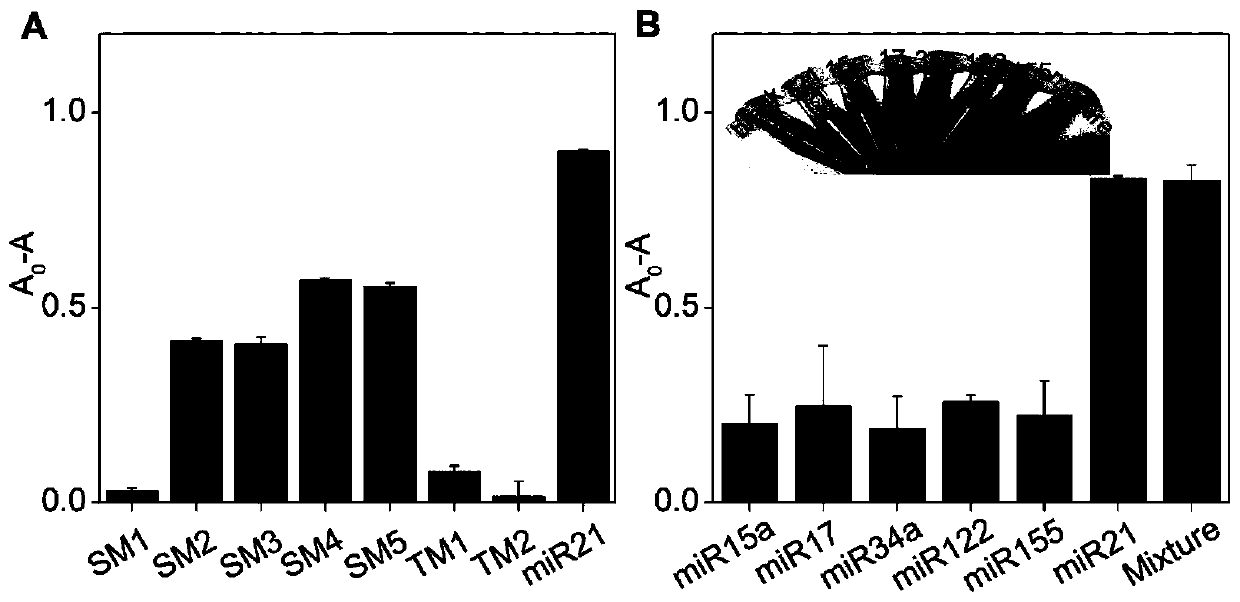
The invention discloses a detection method for the visual recognition of microRNA (Ribonucleic Acid) on the basis of G-quadruplex probe structure unwinding. A signal attenuation method constructed bycombining catalytic characteristic of a G-quadruplex combined with heme with the structure change of the G-quadruplex can distinguish miRNAs of single-base mismatch and multibase mismatch, and also can distinguish different miRNA family members. On the basis of signal attenuation, sensitivity still can be 4.5nmol / L. The probe used by the invention is simple in design, and does not relate to a complex reaction process. The detection method has an important meaning for the detection of miRNA.

Description

technical field [0001] The invention relates to the field of nucleic acid detection, in particular to a detection method for visual recognition of microRNA based on the unwinding of the G-quadruplex probe structure. Background technique [0002] microRNA (hereinafter referred to as miRNA) is a kind of non-coding, single-stranded small RNA with a length of about 22-23 nucleotides that widely exists in eukaryotes. The expression levels of miRNA are different in different cells or tissue types, and they are widely involved in different physiological activities. They can regulate cell proliferation, differentiation and apoptosis mainly through complete degradation or translational repression of target genes. As tumor suppressor genes or proto-oncogenes, miRNAs participate in the occurrence and development of tumors by regulating the expression of apoptotic proteins, kinases and other tumor-inducing factors, so abnormal miRNA expression is considered to be a characteristic of tum...

Claims


Application Information

Patent Timeline
IPC IPC(8): C12Q1/6816
CPCC12Q1/6816
Inventor 程靓兰玲刘利
Owner INST OF CHEM CHINESE ACAD OF SCI
Learn More