Optical imaging device and method for mammary gland based on image segmentation and time-frequency information fusion
An image segmentation and optical imaging technology, applied in image analysis, image enhancement, mammography, etc., can solve the problems of low reconstruction accuracy and low reconstruction efficiency, and achieve the effect of improving reconstruction accuracy, narrowing the scope, and reducing the number
- Summary
- Abstract
- Description
- Claims
- Application Information
AI Technical Summary
Problems solved by technology
Method used
Image
Examples
specific Embodiment approach 1
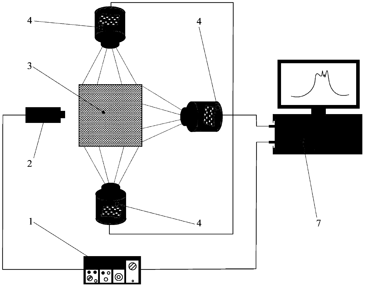
[0040] Specific implementation mode one: combine figure 1 The present embodiment is described. The breast optical imaging device based on image segmentation and time-frequency information fusion provided in this embodiment specifically includes a laser controller 1, a laser head 2, a breast tissue to be measured 3, a data acquisition and processing system 7, and several Microlens array light field camera 4;
[0041] One end of the laser controller 1 is connected to the laser control signal output end of the laser head 2, and the other end of the laser controller 1 is connected to the data acquisition and processing system 7; the signal input end of the data acquisition and processing system 7 is connected with the microlens array light field camera simultaneously 4 is connected to the signal output end; wherein, the plurality of microlens array light field cameras 4 are on the same plane as the laser head 2, and are evenly distributed around the breast tissue 3 to be measured;...
specific Embodiment approach 2
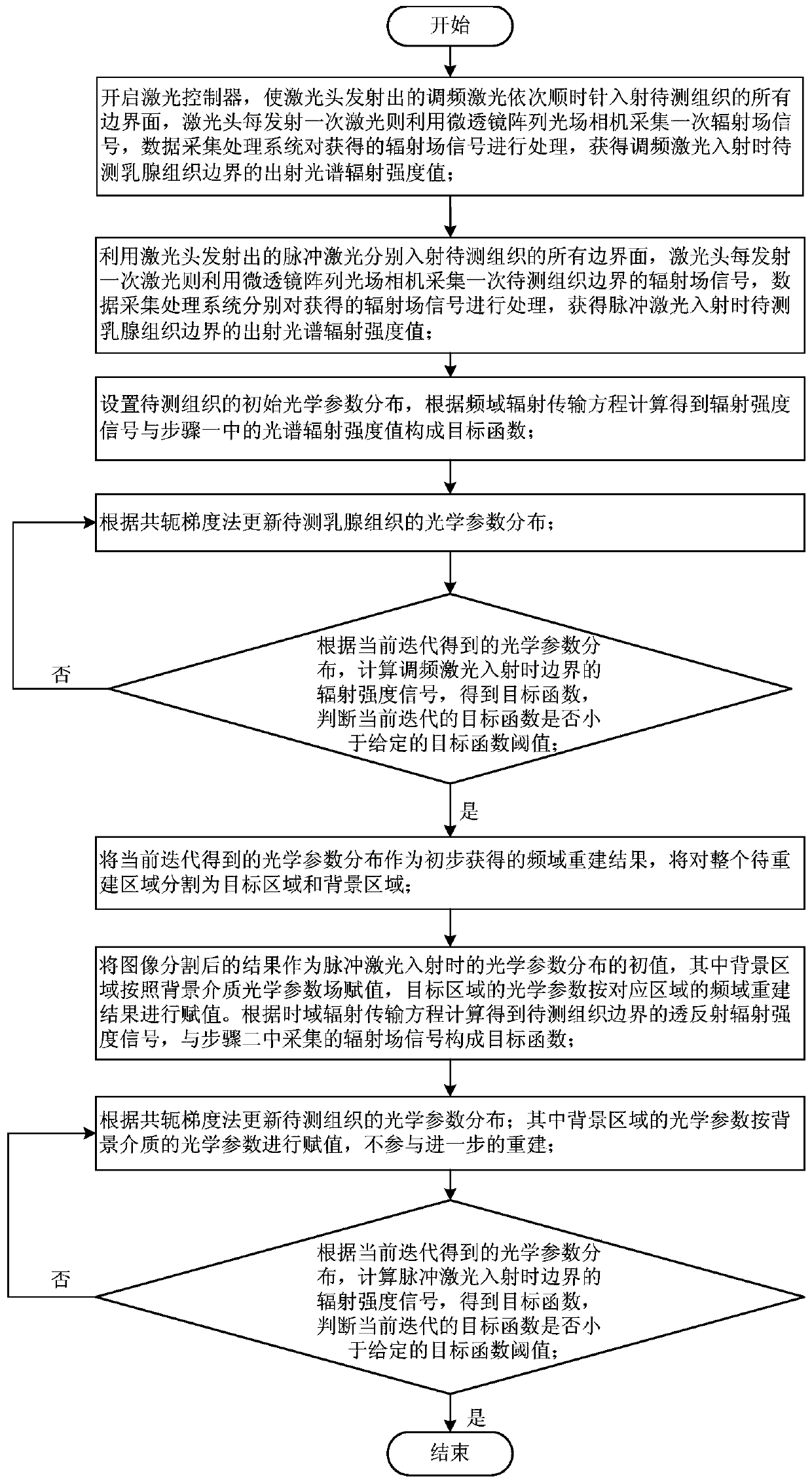
[0042] Specific implementation mode two: combination figure 2 This embodiment will be described. The breast optical imaging method based on image segmentation and time-frequency information fusion provided in this embodiment specifically includes the following steps:
[0043] Step 1. Turn on the laser controller 1, make the frequency-modulated laser emitted by the laser head 2 incident on the breast tissue 3 to be measured (generally in vivo detection and imaging), and then rotate the breast tissue 3 to be measured clockwise to irradiate the frequency-modulated laser The next adjacent boundary surface of the current boundary surface of the breast tissue 3 to be tested is repeatedly rotated until the frequency-modulated laser emitted by the laser head 2 is incident once from each boundary surface of the breast tissue 3 to be measured;
[0044] Every time the laser head 2 emits a frequency-modulated laser, each microlens array light field camera 4 is used to collect the radiati...
specific Embodiment approach 3
[0068] Specific embodiment three: the difference between this embodiment and specific embodiment two is that the optical parameter distribution μ of the breast tissue to be measured includes an absorption coefficient field μ a and the scattering coefficient field μ s Two-part parameters, and the two-part parameter fields are reconstructed at the same time.
[0069] Other steps and parameters are the same as in the second embodiment.
PUM
 Abstract
Description
Claims
Application Information
 - Generate Ideas
- Intellectual Property
- Life Sciences
- Materials
- Tech Scout
- Unparalleled Data Quality
- Higher Quality Content
- 60% Fewer Hallucinations
Browse by: Latest US Patents, China's latest patents, Technical Efficacy Thesaurus, Application Domain, Technology Topic, Popular Technical Reports.
© 2025 PatSnap. All rights reserved.Legal|Privacy policy|Modern Slavery Act Transparency Statement|Sitemap|About US| Contact US: help@patsnap.com


