
All-digital mammary gland imaging image radiomics method based on deep learning

A technology of breast imaging and deep learning, applied in the field of radiomics for the identification of benign and malignant breast tumors, can solve the problem of insufficient information coverage of quantitative image features

Inactive Publication Date: 2018-07-20
SOUTHERN MEDICAL UNIVERSITY
View PDF4 Cites 5 Cited by
  • Summary
  • Abstract
  • Description
  • Claims
  • Application Information

AI Technical Summary

Problems solved by technology

[0006] Therefore, in view of the lack of quantitative image features covered by radiomics in the identification of breast tumors, a deep learning-based radiomics method for all-digital breast imaging images is provided to obtain the abstract features of tumor levels and add them to the radiomics classifier learning in order to overcome the limitations of existing traditional features

Method used


Image

  • All-digital mammary gland imaging image radiomics method based on deep learning
  • All-digital mammary gland imaging image radiomics method based on deep learning

Examples


Embodiment 1

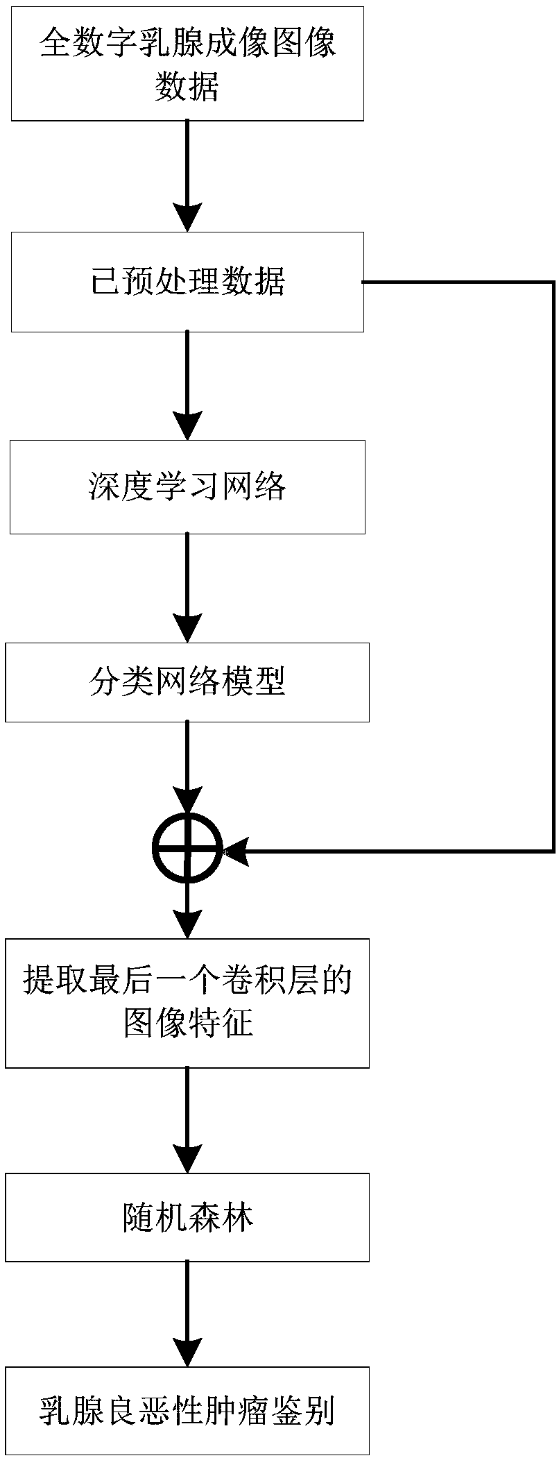
[0039] Such as Figure 1-2 As shown, a radiomics method of full digital breast imaging images based on deep learning, the specific steps are as follows:

[0040] S1. Acquire full digital mammography image data μ datset .

[0041] S2. Through the full digital mammography image data μ datset Perform preprocessing to obtain preprocessed data μ hdf5 .

[0042] The specific steps in step S2 are as follows:

[0043] S21. Perform full digital mammography image data μ datset Carry out segmentation to obtain the divided data μ patch .

[0044] In step S21, the full digital breast imaging image data μ datset Segment the lesion point as the center, and segment the segmented data μ with a size of 572×572 patch .

[0045] S22. For data μ patch Carry out the amplification operation to obtain n amplification data μ 1 expand ,...,μ i expand ,...,μ n expand , where 1≤i≤n, i and n are both integers.

[0046] The amplification operation is to divide the data μ patch Perform i ...


PUM


Abstract

An all-digital mammary gland imaging image radiomics method based on deep learning includes the following specific steps: obtaining all-digital mammary gland imaging image data [mu]<datset>, preprocessing the all-digital mammary gland imaging image data [mu]<datset> to obtain preprocessed data [mu]<hdf5>, inputting the preprocessed data [mu]<hdf5> to a deep learning network Alexnet to perform training, establishing a classification network model M<alexnet>, inputting the preprocessed data [mu]<hdf5> to the classification network model M<alexnet> to perform feature extraction, obtaining a high-dimensional feature vector FeatureMap, inputting the high-dimensional feature vector FeatureMap to a random forest RF to perform training to obtain a high-performance tumor identification classifier. The method provided by the invention uses a deep learning network Alexnet framework to extract image features for identifying a tumor, and combined with radiomics, the random forest is adopted to learn the extracted features, thereby realizing research of all-digital mammary gland imaging image radiomics based on deep learning.

Description

technical field [0001] The invention relates to the technical field of radiomics for the identification of benign and malignant breast tumors, in particular to a radiomics method for full digital breast imaging images based on deep learning. Background technique [0002] In recent years, the organic integration of big data technology and medical image-assisted diagnosis has produced a new radiomics method, which can effectively solve the problem of difficult quantitative evaluation of tumor abnormalities by extracting high-dimensional quantitative features from medical images to quantify major diseases such as tumors. Qualitative questions with important clinical value. [0003] The processing flow of radiomics is summarized into the following parts: (1) acquisition of image data; (2) calibration of tumor area; (3) segmentation of tumor area; (4) extraction and quantification of features; (5) feature extraction; (6) Train and test the classifier. [0004] At present, there...

Claims


Application Information

Patent Timeline
Patent Type & Authority Applications(China)
IPC IPC(8): G06K9/62G06N99/00G06T7/11
CPCG06N20/00G06T7/11G06T2207/10081G06T2207/20081G06T2207/30096G06F18/24
Inventor 边兆英梁翠霞曾栋黄静马建华
Owner SOUTHERN MEDICAL UNIVERSITY
Learn More