ST2 detection kit based on bimolecular fluorescence complementation technology and preparation and use method thereof

A detection kit and bimolecular technology, applied in the field of bimolecular fluorescence complementation, can solve the problems of poor repeatability, heterogeneous reaction, large intra-batch and inter-batch variation, etc.

Inactive Publication Date: 2018-07-10
NANJING TZONE BIOLOGICAL SCI & TECH
View PDF1 Cites 2 Cited by
  • Summary
  • Abstract
  • Description
  • Claims
  • Application Information

AI Technical Summary

Problems solved by technology

The enzyme-linked immunosorbent assay uses horseradish peroxidase or alkaline phosphatase to label antibodies, and catalyzes the color change of the substrate. Low, currently mainly used in infectious disease screening and other projects that require low detection sensitivity; immunochromatography has the advantages of simple operation and fast detection speed, but also has low sensitivity, unstable reagents, and poor repeatability , the disadvantage of being difficult to quantify
Chemiluminescence immunoassay has the advantages of simple operation, fast detection speed, and high-throughput detection, but it also has the disadvantages of heterogeneous reaction, long detection time, and large intra-assay and inter-assay variability.

Method used


Image

  • ST2 detection kit based on bimolecular fluorescence complementation technology and preparation and use method thereof
  • ST2 detection kit based on bimolecular fluorescence complementation technology and preparation and use method thereof
  • ST2 detection kit based on bimolecular fluorescence complementation technology and preparation and use method thereof

Examples


Embodiment 1

[0027] The anti-ST2 antibody is coupled to the N-terminal fragment of fluorescent protein, taking the yellow fluorescent protein (YFP) 1-154 amino acid fragment YFPN as an example, the specific implementation process is as follows:

[0028] 1) Add 0.1mg of YFPN protein into a centrifuge tube, and prepare with 0.05mol / L pH9.5 carbonate buffer (CB) to dilute the YFPN protein to 1mg / mL.

[0029] 2) Add glutaraldehyde at a final concentration of 1.25% in a fume hood.

[0030] 3) Reaction in a water bath at 37°C for 2 hours.

[0031] 4) Dialyze with desalting column Sephadex G-25 or 0.05mol / LpH9.5 CB to remove excess glutaraldehyde.

[0032] 5) Take 0.1mg of anti-ST2 antibody, prepare 1mg / mL antibody with 0.05mol / L pH9.5 CB, and mix the activated YFPN protein with the antibody.

[0033] 6) React overnight at 4°C.

[0034] 7) Blocking: add 50 μL of 0.2 mol / L lysine solution, and block for 2 hours at room temperature to block residual aldehyde groups and terminate the reaction.

...

Embodiment 2

[0038] The anti-ST2 antibody is coupled to the fluorescent protein C-terminal fragment, taking the yellow fluorescent protein (YFP) 155-238 amino acid fragment YFPC as an example, the specific implementation process is as follows:

[0039] 1) Add 0.1mg of YFPC protein into a centrifuge tube, and prepare with 0.05mol / L pH9.5 carbonate buffer (CB) to dilute the YFPC protein to 1mg / mL.

[0040] 2) Add glutaraldehyde at a final concentration of 1.25% in a fume hood.

[0041] 3) Reaction in a water bath at 37°C for 2 hours.

[0042] 4) Dialyze with desalting column Sephadex G-25 or 0.05mol / LpH9.5 CB to remove excess glutaraldehyde.

[0043] 5) Take 0.1mg of anti-ST2 antibody, prepare 1mg / mL antibody with 0.05mol / L pH9.5 CB, and mix the activated YFPC protein and antibody.

[0044] 6) React overnight at 4°C.

[0045] 7) Blocking: add 50 μL of 0.2 mol / L lysine solution, and block for 2 hours at room temperature to block residual aldehyde groups and terminate the reaction.

[0046...

Embodiment 3

[0049] The main components of the kit:

[0050] 1) N-terminal fragment of fluorescent protein coupled with anti-ST2 antibody;

[0051] 2) Anti-ST2 antibody-coupled fluorescent protein C-terminal fragment.


PUM


Abstract

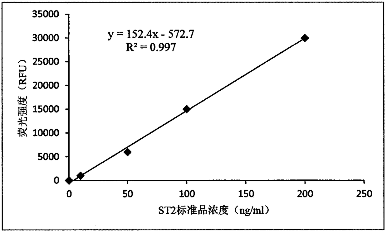
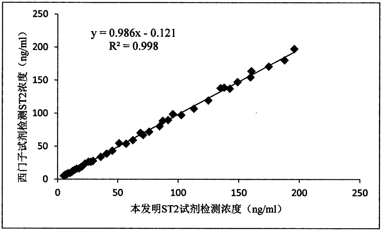
The invention provides an ST2 detection kit based on a bimolecular fluorescence complementation technology and a preparation and use method thereof. The kit includes a fluorescent protein N-end segment resistant to ST2 antibody coupling and a fluorescent protein C-end segment resistant to ST2 antibody coupling. The invention also discloses the preparation method of the ST2 diagnosis kit based on the bimolecular fluorescence complementation technology. The preparation method includes preparation of the fluorescent protein N-end segment resistant to ST2 antibody coupling and preparation of the fluorescent protein C-end segment resistant to ST2 antibody coupling. Finally, the invention also discloses the use method of the kit. The kit has the advantages of being easy to operate, wide in linear range, excellent in specificity, free of cleaning, high in accuracy and the like and can provide convenience for clinical detection and usage; the kit is used for predicting unfavorable prognosis cardiovascular events happening to patients suffering from acute myocardial infarction (AMI) and monitoring the capacity of evaluating the coronary artery disease degree; by means of the kit, the clinical evaluation and risk stratification accuracy of clinicians on AMI patients or AMI high-risk groups are improved, and therefore the kit has a considerable market value.

Description

technical field [0001] The invention relates to a bimolecular fluorescence complementary technology used for in vitro immunodiagnosis to detect the content of ST2 in a human body, belonging to the field of disease diagnosis and detection. Background technique [0002] Growth stimulation expressed gene 2 protein (growth stimulation expressed gene 2, ST2) is a member of the IL-1 receptor superfamily, first discovered by Tominaga in 1989, it has long been considered as an orphan receptor associated with inflammation and immune diseases , and discovered its specific ligand IL-33 in 2005, so the research on ST2 expanded to a new field. Human ST2 gene is located on chromosome 2q12, about 40KD, expressed in mast cells, macrophages, activated helper T cells 2 (Th2), cardiomyocytes and cardiac fibroblasts. [0003] There are 4 transcripts of ST2 gene, among which the 2 most important isoforms are transmodel ST2 (ST2L) and soluble ST2 (sST2), which are formed by promoter alternative ...

Claims


Application Information

Patent Timeline
Patent Type & Authority Applications(China)
IPC IPC(8): G01N33/68G01N33/544G01N33/533
CPCG01N33/533G01N33/544G01N33/6869G01N2333/545
Inventor 徐林
Owner NANJING TZONE BIOLOGICAL SCI & TECH
Why Eureka
Try Eureka