Method for rapidly preparing intraoperative individual covered stent for treating aortic arch dissection
An aortic arch and stent-graft technology, applied in the field of biomedical engineering, can solve the problems of inability to popularize in grass-roots hospitals, many complications, and time-consuming, and achieve the advantages of shortening the time of deep hypothermic circulatory arrest, reducing serious complications, and shortening operation time. Effect
- Summary
- Abstract
- Description
- Claims
- Application Information
AI Technical Summary
Problems solved by technology
Method used
Image
Examples
Embodiment 1
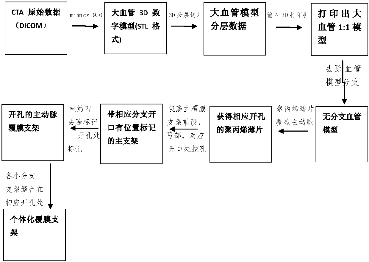
[0039] A process for quickly preparing an intraoperative individualized stent graft for the treatment of aortic arch dissection is as follows: figure 1 Shown:
[0040] 1. Model reconstruction
[0041] Equipment used Discovery CT 750HD (GE Company, the United States) 64 multi-slice spiral CT scanner. The patient was placed in the supine position and breath-holding. First, a non-enhanced CT sequence (0.625mm slice thickness) was obtained. A 22G venous indwelling needle was punctured into the cubital vein, and iohexol (onepaque, containing Iodine 350mg I / mL) 100mL, monitor the peak value of the drug, and delay 6-8s after reaching the peak value. The target area starts from the root of the neck and goes down to the pubic symphysis. The scanned data are exported in DICOM format and imported into Mimics 19.0 software for 3D reconstruction of the aortic arch dissection. The extraction range includes the entire aorta. The surface of the model is smooth and modified, and exported in ...
Embodiment 2
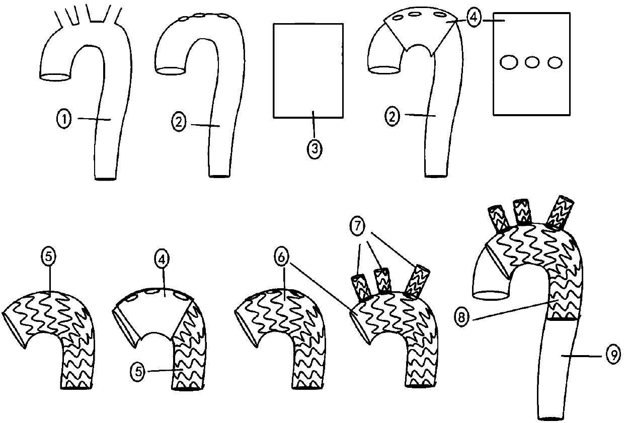
[0049] A process for quickly preparing an intraoperative individualized stent graft for the treatment of aortic arch dissection is as follows: figure 1 Shown:
[0050] 1. Model reconstruction
[0051] Equipment used Discovery CT 750HD (GE Company, the United States) 64 multi-slice spiral CT scanner. The patient was placed in the supine position and breath-holding. First, a non-enhanced CT sequence (0.625mm slice thickness) was obtained. A 22G venous indwelling needle was punctured into the cubital vein, and iohexol (onepaque, containing Iodine 350mg I / mL) 100mL, monitor the peak value of the drug, and delay 6-8s after reaching the peak value. The target area starts from the root of the neck and goes down to the pubic symphysis. The scanned data are exported in DICOM format and imported into Mimics 19.0 software for 3D reconstruction of the aortic arch dissection. The extraction range includes the entire aorta. The surface of the model is smooth and modified, and exported in ...
Embodiment 3
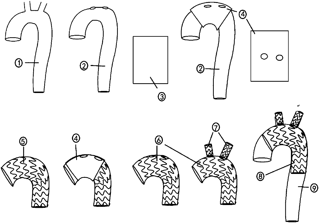
[0059] A process for quickly preparing an intraoperative individualized stent graft for the treatment of aortic arch dissection is as follows: figure 1 Shown:
[0060] 1. Model reconstruction
[0061] Equipment used Discovery CT 750HD (GE Company, the United States) 64 multi-slice spiral CT scanner. The patient was placed in the supine position and breath-holding. First, a non-enhanced CT sequence (0.625mm slice thickness) was obtained. A 22G venous indwelling needle was punctured into the cubital vein, and iohexol (onepaque, containing Iodine 350mg I / mL) 100mL, monitor the peak value of the drug, and delay 6-8s after reaching the peak value. The target area starts from the root of the neck and goes down to the pubic symphysis. The scanned data are exported in DICOM format and imported into Mimics 19.0 software for 3D reconstruction of the aortic arch dissection. The extraction range includes the entire aorta. The surface of the model is smooth and modified, and exported in ...
PUM
 Abstract
Description
Claims
Application Information
 - Generate Ideas
- Intellectual Property
- Life Sciences
- Materials
- Tech Scout
- Unparalleled Data Quality
- Higher Quality Content
- 60% Fewer Hallucinations
Browse by: Latest US Patents, China's latest patents, Technical Efficacy Thesaurus, Application Domain, Technology Topic, Popular Technical Reports.
© 2025 PatSnap. All rights reserved.Legal|Privacy policy|Modern Slavery Act Transparency Statement|Sitemap|About US| Contact US: help@patsnap.com


