
Staining composition for staining an ophthalmic membrane

a technology of ophthalmic membrane and staining composition, which is applied in the direction of luminescence/biological staining preparation, drug composition, biocide, etc., can solve the problems of difficult removal of membranes, retinal damage, and difficulty in creating continuous curvilinear capsulorhexis (ccc) in eyes with white mature cataracts

Active Publication Date: 2010-06-08
KYUSHU UNIV
View PDF6 Cites 8 Cited by
  • Summary
  • Abstract
  • Description
  • Claims
  • Application Information

AI Technical Summary

Benefits of technology

[0083]According to one preferred embodiment of the present invention, said staining composition can be used as a surgical adjuvant in ophthalmic surgeries related to ophthalmic diseases, such as, for example, vitreo-retinal disease such as Macular hole, retinal detachment due to hymyopic macular hole, epiretinal membrane, proliferative diabetic retinopathy, diabetic macular edema, and proliferative vitreoretinopathy, and specific cataracts such as hypermature cataract, and congenital cataract, and split-thickness corneal transplantation, etc. According to the staining composition of the present invention, it becomes possible to more clearly verify difficult-to-see ophthalmic membranes, and improve safety during surgical operations.

Problems solved by technology

It is now widely recognized that without surgical adjuvant it is extremely difficult to remove the membranes due to the poor visibility of the ILM and ERMS.
In particular, Indocyanine green (ICG) and Trypan Blue (TB) staining have greatly facilitated ILM and ERMS peeling in various vitreo-retinal diseases and as a result, this technique is now widely accepted by many surgeons However, numerous reports have recently emerged regarding retinal damage caused by intravitreal injections of ICG and TB both in experimental and clinical use.
Further, creating a continuous curvilinear capsulorhexis (CCC) in eyes with a white mature cataract can be challenging as it is difficult to distinguish the anterior capsule from the underlying white cortex.
Poor visualization of the capsule tends to result in an incomplete or inadequate CCC that could cause a subsequent capsular tear, vitreous loss and intraocular lens (IOL) dislocation.
However, trypan blue has been reported to be toxic to corneal endothelium in vitro in severe conditions.
However, the inventors reported the potential toxicity of ICG to retinal cells in 2002, and recently, the toxicity of ICG has been reported in retinal pigment epithelium, ganglion cells, and photoreceptors.
Such doubts developed due to reports of the possibility of toxicity, teratogenicity and so on to the retinal cells.
Furthermore, there were technical problems such as the inability to get a satisfactory dye at lower concentrations and complicated staining methods.
Thus, such problems made ophthalmic surgery an even more difficult matter.

Method used


Image

  • Staining composition for staining an ophthalmic membrane
  • Staining composition for staining an ophthalmic membrane
  • Staining composition for staining an ophthalmic membrane

Examples


example 1

Screening Various Dyes as a Candidate Focusing on their Safety and Ability to Stain Membranes

[0202]In order to provide a staining composition for achieving such aims, all dyes, including dyes currently not being utilized for organisms were used, and they were then screened for candidate dyes. The dyes screened are Evans blue, Acid blue 9, Fast green, Brilliant Blue FCF, Indigo-carmine, methyl blue, methylene blue, Brilliant Blue G (BBG). Additionally, as the screening conditions, safety and staining-affinity were especially noted.

[0203]Among some of the leading candidates, those that showed superiority in safety but inability to dye the target object, or in the reverse, those that showed good staining but virulence to the retina, were removed from candidacy.

[0204]Safety assessments were then conducted, using rats, of the narrowed down candidate dyes. Based on higher staining-affinity and safety, BBG became the final candidate.

[0205]Next, in order to examine the optimal conditions of...

example 2

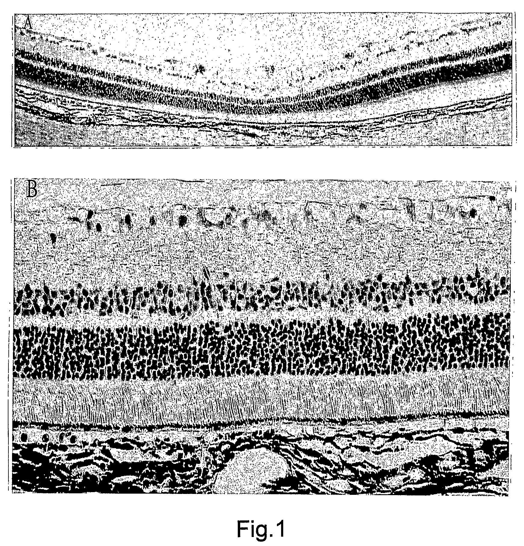
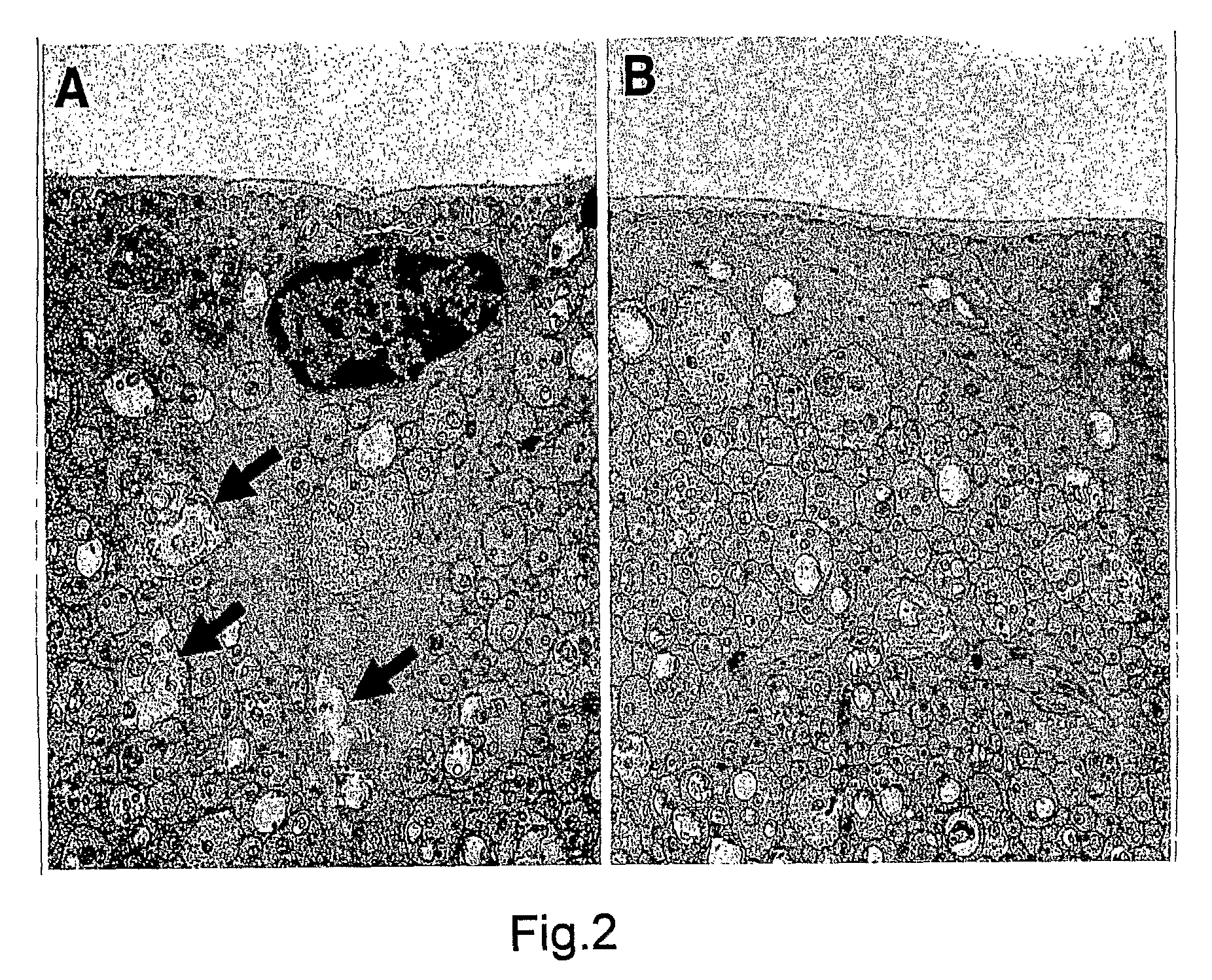
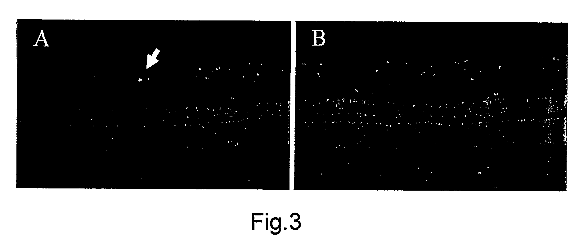
2. Effects of the Intravitreous BBG on the Retina

[0206]All procedures conformed to the Association for Research in Vision and Opthalmology (ARVO) statement for the Use of Animals in Ophthalmic and Vision Research and the guidelines for animal care at Kyushu University, Fukuoka, Japan.

2.1 Characterization of the BBG Solution

[0207]BBG is also known as Acid Blue 90 and Coomassie Brilliant Blue G. Aside from our previous publication, there are no reports investigating the toxicity BBG for ophthalmic use. Table 1 shows the osmolarity and pH of the BBG solution at different concentrations. The osmolarity and pH of BBG was found to be similar to those of intraocular irrigating solutions.

[0208]

TABLE 1solutionOsmolarity (mOsm / KgH20)pHcontrol2987.33BBG 10 mg / ml3107.41BBG 1.0 mg / ml3007.42BBG 0.1 mg / ml2987.41BBG 0.01 mg / ml2987.41saline2857.4BSS3057.1plus (Registeredtrademark)Control shows OPEGUARD ® - MA.

2.2. Surgical Procedure for Intravitreal BBG in Rat Eyes

[0209]Brown Norway rats (n=78 males...

example 3

3. Interventional Clinical Case Series of BBG as a Potential Staining Solution for Membrane Peeling

[0222]Indocyanine green (ICG) and Trypan Blue (TB) staining have greatly facilitated ILM peeling in various vitreo-retinal diseases. However, numerous reports have recently emerged regarding retinal damage caused by ICG and TB both in experimental and clinical use.

[0223]From the preclinical study, BBG was selected as a candidate dye for ILM staining. This is apparently the first clinical investigation of BBG in human eyes. This is the first known clinical investigation of BBG in human eyes.

3.1. The Use of BBG in ILM Staining and the Effects and Safety in Human Eyes (the First Attempt)

[0224]Sixteen eyes of 16 patients with a macular hole (MH) and epiretinal membranes (ERMS) were recruited from the outpatient unit of Kyushu University Hospital (Fukuoka, Japan) between August and September 2004. Patients with ocular diseases such as glaucoma, uveitis, and corneal disorder were excluded. C...


PUM

PropertyMeasurementUnit
concentrationaaaaaaaaaa

Abstract

A staining composition for staining an ophthalmic membrane when performing the ophthalmic membrane removal, wherein the staining composition comprises a Brilliant Blue G (BBG) derivative as a primary component.

Description

PRIORITY FILING[0001]This application claims priority to U.S. Provisional Application Nos. 60 / 633,592 filed on Dec. 6, 2004, and 60 / 647,504 filed on Jan. 27, 2005, the contents of which are incorporated herein by reference in their entirety.FIELD OF THE INVENTION[0002]The present invention relates to a staining composition for staining an ophthalmic membrane. More specifically, the present invention relates to the staining composition used as an adjuvant for staining the ophthalmic membrane specifically to remove the membrane.BACKGROUND OF THE INVENTION[0003]The staining of the internal limiting membrane (ILM) is one of the important developments of surgery for such vitreo-retinal diseases as macular hole and epiretinal membranes (ERMS) (Indocyanine green-assisted peeling of the retinal internal limiting membrane. Burk S E et al. Opthalmology. 2000; 107:2010-2014). It is now widely recognized that without surgical adjuvant it is extremely difficult to remove the membranes due to the...

Claims


Application Information

Patent Timeline
Patent Type & Authority Patents(United States)
IPC IPC(8): A61K49/00A61K31/135
CPCA61K49/006A61K31/185A61P27/02
Inventor ENAIDA, HIROSHIHISATOMI, TOSHIOISHIBASHI, TATSUROKAGIMOTO, TADAHISAHATA, YASUAKI
Owner KYUSHU UNIV
Learn More