
Superfine examination gastroscope

A gastroscope and introduction tube technology, which is applied to the field of gastroscopes with ultra-fine diameters, can solve the problems of inability to popularize physical examination screening, residual lesions, and difficulty in manipulation, and achieve the effects of obvious inspection costs, small and reasonable structure, and controllability.

Pending Publication Date: 2017-12-12
胡广禄
View PDF13 Cites 2 Cited by
  • Summary
  • Abstract
  • Description
  • Claims
  • Application Information

AI Technical Summary

Problems solved by technology

However, in clinical use, it was found that because the stay and shooting angle of the capsule gastroscope depend on the dynamics of the swallower’s gastrointestinal tract, the controllability is poor and the manipulation is difficult, and the field of view cannot fully cover the remaining risk of dead-angle lesions.
[0006] At the same time, both kinds of examinations are expensive due to the high cost of equipment (600-700 yuan for painless gastroscopy, 3000-5000 yuan for ordinary capsule gastroscopy, and higher price for magnetically controlled capsule gastroscopy). The cost of the examination, as a physical examination screening, cannot be popularized, and early detection and early intervention of digestive tract diseases cannot be implemented. The incidence of gastric cancer and esophageal cancer in China ranks in the middle of the world, but the proportion of advanced gastric and esophageal cancer and the mortality rate are among the top in the world

Method used


Image

  • Superfine examination gastroscope
  • Superfine examination gastroscope
  • Superfine examination gastroscope

Examples


Embodiment Construction

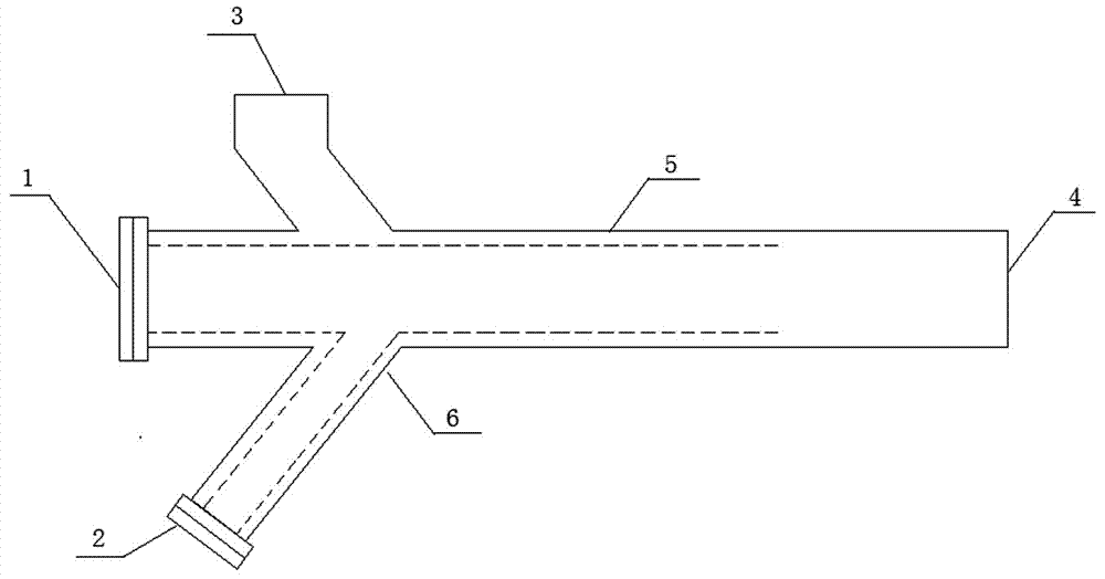
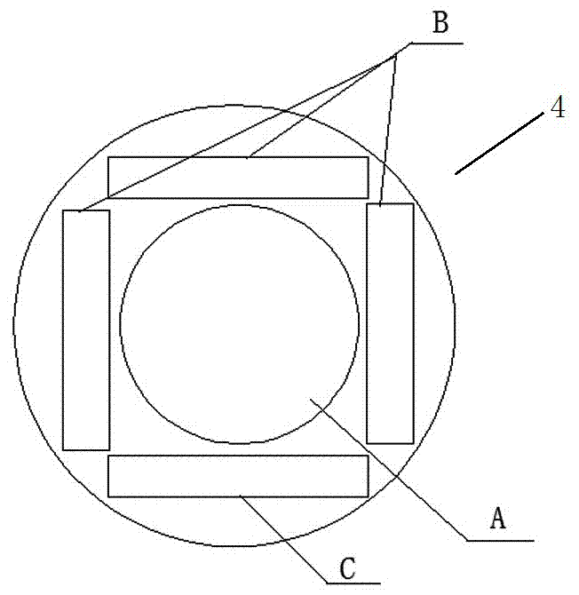
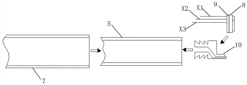
[0030] Refer to attached Figure 1~5 , the ultra-fine inspection gastroscope of the present invention comprises main body, and described main body is divided into importing part, operating part and guide wire 11, and importing part adopts PVC material as introducing pipe body 5, and the length of introducing pipe body 5 is 1030 millimeters, and outer diameter is 2 mm, with an inner diameter of 1.2 mm; an illumination and image acquisition unit is provided at the front end of the introduction tube body 5, and the rear part of the introduction tube body 5 is connected with the operation part; the operation part adopts a three-way body 6, which is divided into two operation ports and one The fast electrical interface 3, the first operating port 1 in the middle of the three-way body 6 is the guide wire entrance, and the guide wire 11 is inserted into the introduction part through the guide wire entrance. The gap between the tube body 5 and the guide wire 11 is used to transport wa...


PUM

PropertyMeasurementUnit
Lengthaaaaaaaaaa
Outer diameteraaaaaaaaaa
Inner diameteraaaaaaaaaa

Abstract

The invention discloses a superfine examination gastroscope, and relates to an esophagus and stomach examination instrument, in particular to the gastroscope of which the diameter is superfine. The superfine examination gastroscope comprises a body, the body is divided into a guide-in part, an operating part and a guiding wire, a guide-in pipe body of the guide-in part is made from a PVC material, the length of the guide-in pipe body is 1,030 millimeters, the outer diameter is 2 millimeters, and the inner diameter is 1.2 millimeters; an illumination and image collection unit is arranged at the front end portion of the guide-in pipe body, and the rear portion of the guide-in pipe body is connected with the operating part; a three-way body is adopted in the operating part, and is divided into two operating openings and one quick electrical interface, the first operating opening in the middle of the three-way body serves as a guiding wire inlet, and the guiding wire is inserted into the guide-in part through the guiding wire inlet; gaps between the guide-in pipe body and the guiding wire are used for delivering water and gas, and a water and gas spraying opening is formed in the front end of the guide-in pipe body; an XLR interface is adopted as the quick electrical interface of the three-way body; the second operating interface of the lateral part of the three-way body is connected with a water / gas unit. The superfine examination gastroscope is exquisite and reasonable, can be accepted by patients, and is highly worthy of popularization.

Description

technical field [0001] The invention relates to an ultrafine examination gastroscope, which relates to an esophagus and stomach inspection instrument, in particular to a gastroscope with ultrafine diameter. Background technique [0002] In modern times, due to changes in people's lifestyles and the acceleration of the pace of life, fast food is favored by consumers. Gastrointestinal diseases occur frequently, and gastric cancer is a disease with a high incidence. [0003] Existing esophagus and gastric cavity inspection adopts two methods: [0004] 1. Traditional gastroscopy requires a thicker introduction tube (the outer diameter of the conventional main tube is 9.8 mm), which causes severe discomfort to the examinee during the examination and requires anesthesia; many patients are unable to tolerate it or are unwilling to accept traditional gastroscopy due to fear. The endoscopic insertion examination, thus delaying the early diagnosis and timely treatment of the disease;...

Claims


Application Information

Patent Timeline
IPC IPC(8): A61B1/273A61B1/012A61B1/05A61B1/06
CPCA61B1/00119A61B1/012A61B1/05A61B1/0684A61B1/273
Inventor 胡歆迪胡广禄
Owner 胡广禄
Learn More