Oocyte separation and extraction method based on duck embryo
An oocyte and extraction method technology, applied in the field of oocyte separation and extraction, can solve the problems of affecting research, time-consuming, affecting oocyte extraction, etc., so as to reduce cell contamination, reduce test costs, and achieve fast and efficient acquisition. Effect
- Summary
- Abstract
- Description
- Claims
- Application Information
AI Technical Summary
Problems solved by technology
Method used
Image
Examples
Embodiment 1
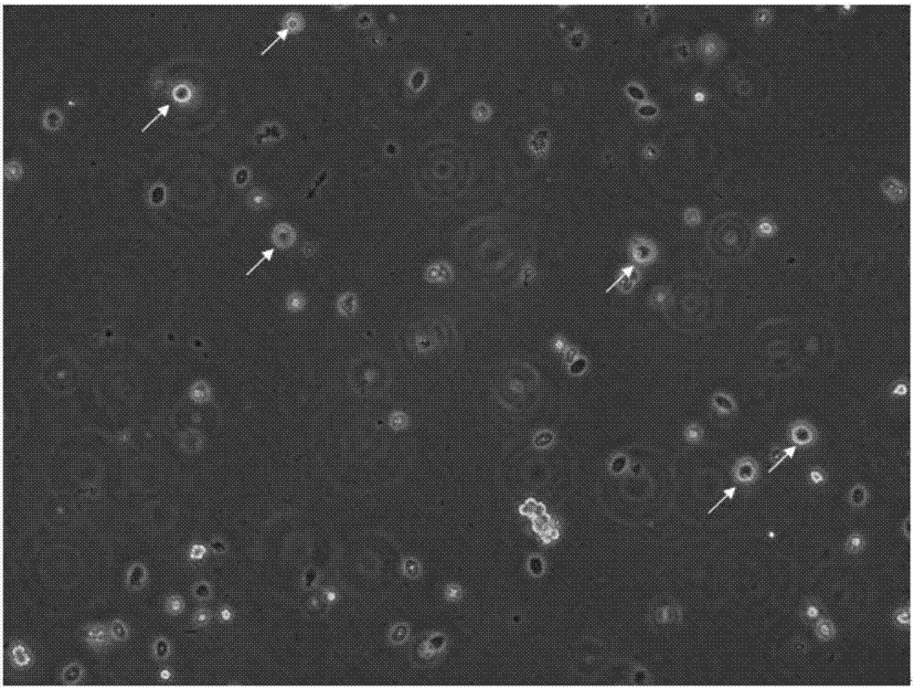
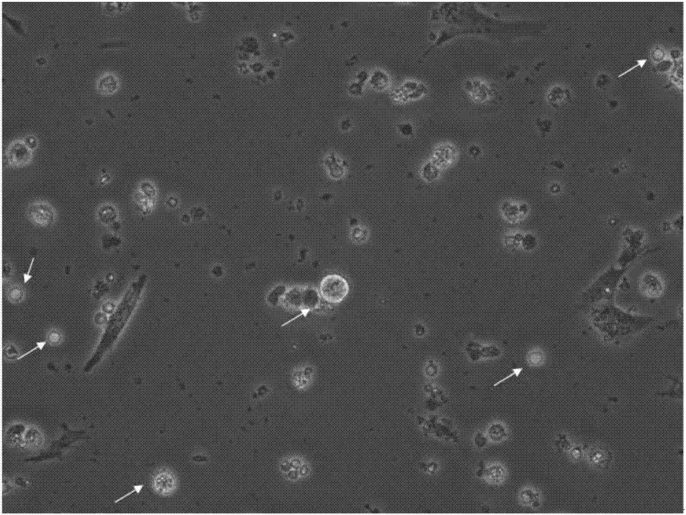
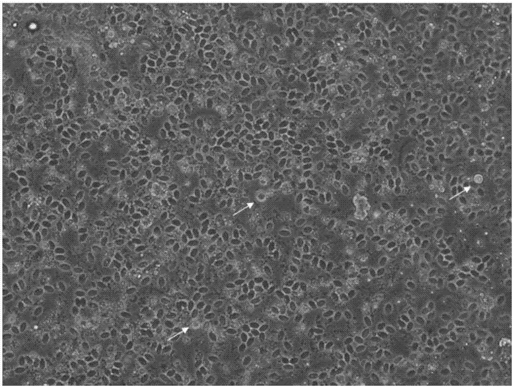
[0031] A method for separating and extracting oocytes based on duck embryos, comprising the following steps:
[0032] Step 1, select 4 ovary tissues of duck embryos at 25 embryonic age, place the ovary tissues in sterilized PBS, and cut the ovary tissues into pieces with ophthalmic scissors;
[0033] Step 2. Add mixed enzyme digestion solution to the shredded ovarian tissue fragments (each 10ml digestion solution contains 5 mg of collagenase II, protease, and hyaluronidase each), and place the ovarian tissue fragments and mixed enzyme digestion solution in a fine mouth In the digestion bottle, digest on a shaker for 45min, 37°C, 190r / min;
[0034] Step 3. Blow and beat the ovarian tissue fragments with a pipette gun, add 10% fetal bovine serum to stop the digestion, and filter through a 200-mesh cell sieve;
[0035] Step 4. Collect the filtrate and centrifuge for 3.5 minutes at a rotational speed of 1200r / min; discard the filtrate, add cell culture medium to the precipitated ...
Embodiment 2
[0038] A method for separating and extracting oocytes based on duck embryos, comprising the following steps:
[0039] Step 1. Select 6 ovarian tissues of duck embryos with an embryonic age of 24 days, place the ovarian tissues in sterilized PBS, and cut the ovarian tissues into pieces with ophthalmic scissors;
[0040] Step 2. Add mixed enzyme digestion solution (each 10ml digestion solution contains collagenase II, protease, and hyaluronidase 4mg) to the shredded ovarian tissue fragments, and place the ovarian tissue fragments and mixed enzyme digestion solution in a fine mouth In the digestion bottle, digest on a shaker for 50min, 37°C, 170r / min;
[0041] Step 3. Blow and beat the ovarian tissue fragments with a pipette gun, add 10% fetal bovine serum to stop the digestion, and filter through a 200-mesh cell sieve;
[0042] Step 4. Collect the filtrate and centrifuge for 3 minutes at a rotational speed of 1500r / min; discard the filtrate, add cell culture medium to the preci...
Embodiment 3
[0045] A method for separating and extracting oocytes based on duck embryos, comprising the following steps:
[0046] Step 1. Select 5 ovarian tissues of duck embryos with an embryonic age of 26 days, place the ovarian tissues in sterilized PBS, and cut the ovarian tissues into pieces with ophthalmic scissors;
[0047]Step 2. Add mixed enzyme digestion solution (each 10ml digestion solution contains collagenase II, protease, and hyaluronidase 3 mg) to the shredded ovarian tissue fragments, and place the ovarian tissue fragments and mixed enzyme digestion solution in a fine mouth In the digestion bottle, digest on a shaker for 60min, 37°C, 180r / min;
[0048] Step 3. Blow and beat the ovarian tissue fragments with a pipette gun, add 10% fetal bovine serum to stop the digestion, and filter through a 200-mesh cell sieve;
[0049] Step 4. Collect the filtrate and centrifuge for 4 minutes at a rotational speed of 1000r / min; discard the filtrate, add cell culture medium to the preci...
PUM
 Abstract
Description
Claims
Application Information
 - Generate Ideas
- Intellectual Property
- Life Sciences
- Materials
- Tech Scout
- Unparalleled Data Quality
- Higher Quality Content
- 60% Fewer Hallucinations
Browse by: Latest US Patents, China's latest patents, Technical Efficacy Thesaurus, Application Domain, Technology Topic, Popular Technical Reports.
© 2025 PatSnap. All rights reserved.Legal|Privacy policy|Modern Slavery Act Transparency Statement|Sitemap|About US| Contact US: help@patsnap.com


