Dyeing method for observing collagenous fibers by adopting polarization microscope
A staining method and technology for collagen fibers, which are applied in the field of staining of collagen fibers observed by polarized light microscope, can solve the problems of affecting correct observation and lesion analysis, unclear structure of collagen fibers, weakening of yellow lining dye background, etc., and achieve lining dye background. Vivid color, easy for tissue fibrosis, clear coloring effect
- Summary
- Abstract
- Description
- Claims
- Application Information
AI Technical Summary
Problems solved by technology
Method used
Image
Examples
Embodiment 1
[0034] Tissues were obtained from 42-day-old normal and chronic fibrotic mouse livers and fixed with 10% formaldehyde.
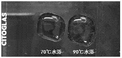
[0035] Picric acid-Sirius red dyeing solution: add 0.1g of picric acid to 1mL Sirius red dyeing solution, after a 90°C water bath, there is insoluble powder at the bottom of the dyeing solution, take the upper solution for later use.
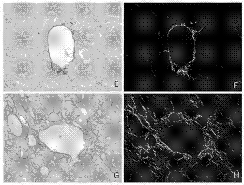
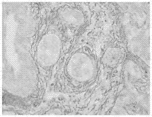
[0036]Staining method: (1) fix the tissue in 10% formaldehyde solution, routinely dehydrate and embed in wax for pathology; (2) paraffin sections with a thickness of 4 μm, routinely dewax to water to obtain tissue sections; (3) drip on the tissue sections Add picric acid-Sirius red staining solution in a water bath at 90°C for 30 minutes; (4) Add 1% w / v acetic acid aqueous solution dropwise and soak for 2 times, each time for 30 seconds; (5) Wash slightly in running water to remove the surface staining (6) Dry in an oven, and fix with neutral resin; (7) Observe under ordinary light microscope and polarized light microscope.
Embodiment 2
[0038] Tissues were obtained from 42-day-old normal and chronic fibrotic mouse livers and fixed with 10% formaldehyde.
[0039] Picric acid-Sirius red dyeing solution: add 0.1g of picric acid to 1mL Sirius red dyeing solution, after a 90°C water bath, there is insoluble powder at the bottom of the dyeing solution, take the upper solution for later use.
[0040] Staining method: (1) fix the tissue in 10% formaldehyde solution, routinely dehydrate and embed in wax for pathology; (2) paraffin sections with a thickness of 4 μm, routinely dewax to water to obtain tissue sections; (3) drip on the tissue sections Add picric acid-Sirius red staining solution in a 70°C water bath for 30 minutes; (4) Add dropwise 1% w / v acetic acid aqueous solution for 2 times, 30s each time; (5) Slightly soak in running water to remove the surface staining of tissue sections (6) Dry in an oven, and fix with neutral resin; (7) Observe under ordinary light microscope and polarized light microscope.
Embodiment 3
[0042] Tissues were obtained from 42-day-old normal and chronic fibrotic mouse livers and fixed with 10% formaldehyde.
[0043] Picric acid-Sirius red dyeing solution: add 0.1g of picric acid to 1ml Sirius red dyeing solution, after 90℃ water bath, there is insoluble powder at the bottom of the dyeing solution, take the upper solution for later use.
[0044] Staining methods: (1) Fix the tissue in 10% formaldehyde solution, routinely dehydrate and make transparent, and embed in wax; (2) Paraffin sections with a thickness of 4 μm are routinely dewaxed to water to obtain tissue sections; (3) Hematoxylin staining for 6 minutes , rinse the tissue sections slightly with running water; (4) stain the tissue sections with picric acid-Sirius red staining solution in a 90°C water bath for 30 min; (5) add 1% w / v acetic acid aqueous solution to soak twice, each time 30s; (6) Slightly immerse in running water to remove the staining solution on the surface of the tissue section; (7) Dry in ...
PUM
 Abstract
Description
Claims
Application Information
 - R&D
- Intellectual Property
- Life Sciences
- Materials
- Tech Scout
- Unparalleled Data Quality
- Higher Quality Content
- 60% Fewer Hallucinations
Browse by: Latest US Patents, China's latest patents, Technical Efficacy Thesaurus, Application Domain, Technology Topic, Popular Technical Reports.
© 2025 PatSnap. All rights reserved.Legal|Privacy policy|Modern Slavery Act Transparency Statement|Sitemap|About US| Contact US: help@patsnap.com


