Scanning human micro-vessel ultra microstructure three-dimensional imaging system

An ultrastructure and three-dimensional imaging technology, applied in medical science, diagnosis with light, sensors, etc., can solve the problems that two-dimensional imaging cannot obtain depth information, two-dimensional imaging cannot meet the requirements, etc., and achieve simple and compact structure, easy operation Convenience and cost reduction effects

Active Publication Date: 2017-08-18
QINGDAO UNIV
View PDF15 Cites 0 Cited by
  • Summary
  • Abstract
  • Description
  • Claims
  • Application Information

AI Technical Summary

Problems solved by technology

[0005] However, the conventional Orthogonal Polarization Spectroscopy (OPS) imaging technology can only present two-dimensional images. Although the analysis of two-dimensional imaging can quantify and digitize micro-blood flow velocity, micro-vessel diameter, and cross-sectional micro-vessel density, Since two-dimensional imaging cannot obtain depth information, two-dimensional imaging cannot meet the requirements for analyzing the microvessel density and microvessel shape in the longitudinal section

Method used


Image

  • Scanning human micro-vessel ultra microstructure three-dimensional imaging system
  • Scanning human micro-vessel ultra microstructure three-dimensional imaging system
  • Scanning human micro-vessel ultra microstructure three-dimensional imaging system

Examples


Embodiment 1

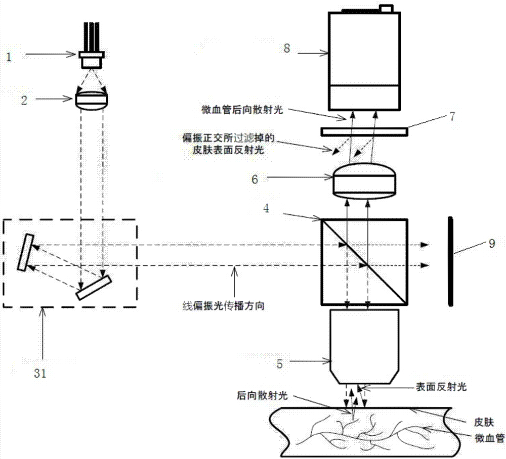
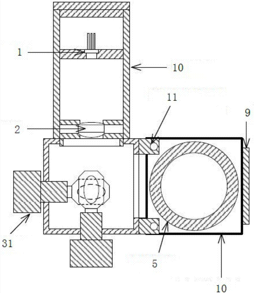
[0045] Such as Figure 2-4 In the scanning microvascular ultrastructure three-dimensional imaging system shown, the scanning module 3 is a two-axis scanning galvanometer system 31 . Specifically, if Figure 5The shown two-axis scanning galvanometer system 31 includes a two-axis galvanometer module connected to the motor control module, the motor control module is divided into an X-axis motor control module and a Y-axis motor control module, and the X-axis motor control module and the Y-axis motor control module The module is connected with the driving module of the vibrating mirror system, and the driving module of the vibrating mirror system is issued a control command by a computer or a microprocessor.

[0046] Specifically, the computer or microprocessor sends instructions to the two-axis scanning galvanometer system 31 to make the light source 1 scan on the plane of the observation area. The calculation coordinate system used by the computer to issue instructions to the ...

Embodiment 2

[0059] When using a scanning 3D microscope such as an ordinary confocal microscope to perform three-dimensional imaging of micro blood flow, due to the slow scanning speed, the real-time imaging effect is poor, so it is difficult to measure the flow of micro blood flow, and it is easy to receive human pulsation and other factors. To realize scanning imaging of micro-blood flow, a device capable of high-speed scanning is needed, and the device can perform non-invasive three-dimensional imaging of micro-blood flow through the skin. Based on the above purpose, a rotating scanning device composed of two upper and lower discs is designed. In this device, the upper disc is the light incident end, embedded with multiple sets of microlenses with different focal lengths; the lower disc is the light output end. There is a light-through small hole at the same position as the focal length of the microlens. From the perspective of mechanical movement, the accuracy and speed of rotation ar...


PUM


Abstract

The invention relates to a scanning human micro-vessel ultra-microstructure three-dimensional imaging system which at least comprises a light source module, a scanning module, a light splitting unit, a scanning lens, an imaging module and an image display module, the light source module is used for emitting emergent light of columnar linear propagation, point-to-point propagation of light rays through the scanning module, one parts of light rays are reflected, other parts of light rays transmit through the light splitting unit, the scanning lens is used for focusing the light rays on one point to irradiate detected object and amplifying micro-blood flow information, the imaging module is used for imaging, and the image display module is used for displaying an image. Micro-vessel ultra-microstructure information in skins is acquired in a light-spot manner through the scanning module by the aid of OPS (optical packet switching) imaging principle, information of imaging light spot brightness is combined in a point-to-point scanning manner, a three-dimensional shape of a micro-vessel ultra-microstructure is restored, point-to-point scanning speed of the scanning module is quite rapid, real-time three-dimensional detection of the micro-vessel ultra-microstructure is achieved, and micro-vessel density, micro-vessel shapes, shapes of micro-vessel endothelial cells and blood cells and the like of a longitudinal section are accurately digitized and quantified.

Description

technical field [0001] The invention relates to a medical device for medical diagnosis by using optical imaging technology, in particular to a scanning three-dimensional imaging system for microvascular ultrastructure of human body. Background technique [0002] Microcirculation refers to the place where blood and tissue cells exchange substances between arterioles and venules. The integrity of the function, shape and metabolism of the microcirculation is an indispensable condition for maintaining the normal function of human organs. Through the study of microcirculation, it is convenient to further understand the special functions of various organs of the human body, and to recognize the pathogenesis of diseases, which is beneficial to disease prevention, diagnosis and treatment. A variety of disease states, including diabetes, hypertension and coronary heart disease, will cause pathological changes in microcirculation, including changes in microvascular diameter, microvas...

Claims


Application Information

Patent Timeline
Patent Type & Authority Applications(China)
IPC IPC(8): A61B5/00
CPCA61B5/0059A61B5/489
Inventor 于綦悦董蒨魏宾夏楠
Owner QINGDAO UNIV
Why Eureka
Try Eureka