Method for preparing autologous beautifying micro-needle preparation and application thereof
A microneedle preparation and autologous technology, which is applied in the direction of medical formula, medical preparations containing active ingredients, drug delivery, etc., can solve the problems of insufficient wrinkle removal effect and poor stain lightening effect, so as to promote microcirculation, Effects of promoting wound healing and improving nutritional status
- Summary
- Abstract
- Description
- Claims
- Application Information
AI Technical Summary
Problems solved by technology
Method used
Image
Examples
example 1
[0039] This example 1 is to isolate autologous PRP and peripheral blood mononuclear cells PBMCs:
[0040] Under sterile conditions, 20ml of peripheral venous blood was collected from the patient with a 20ml syringe, and anticoagulated with heparin sodium. Transfer the anticoagulated peripheral blood to a 50ml centrifuge tube at 400g and centrifuge for 15 minutes. After centrifugation, use a Pasteur pipette to collect the plasma layer and transfer it to a new 15ml centrifuge tube, and suck it up to 1-2 mm above the cell layer. 1500 g of autologous plasma containing platelets for 15 minutes. The upper layer of plasma was collected into a new 15ml centrifuge tube and separated from the lower layer of pellets (retain the bottom 3-5ml of plasma during separation and do not remove it from the tube). The remaining 3-5 ml of plasma and platelet pellet in the tube are resuspended to obtain concentrated PRP. Store the concentrated PRP and the remaining plasma at -80°C; dilute the lowe...
example 2
[0042] This example 2 is to induce fibroblasts, endothelial progenitor cells and immune cells in vitro:
[0043] The isolated PBMCs were resuspended in activated cell initial medium (containing activated cytokine solution), and the cell density was controlled at 1 × 10 6 cells / ml, transfer the cells into a T75 culture flask, shake well, and place them in a CO2 incubator with saturated humidity, 37°C, and 5% CO2 concentration.
[0044] After 2 days of culture, centrifuge at 500g / min for 6 minutes, collect the cells, resuspend the cell pellet with activated cell initial medium (containing activated cytokine solution), transfer it to a T75 culture flask, shake well, and place it in a CO2 incubator The culture was continued at medium saturated humidity, 37°C, and 5% CO2 concentration.
example 3
[0046] Example 3 is the quality inspection of the above-mentioned cultured cells
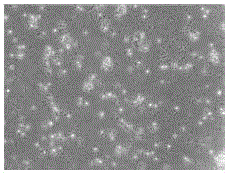
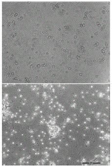
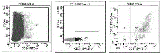
[0047]Morphology, purity, cell phenotype, cell number, viability, sterility, endotoxin, etc. were tested on the cells on the third day of culture. The specific operation includes the following steps: take 0.5ml of cell fluid to count with a cell counter, trypan blue staining to measure the cell viability, and flow cytometry to detect the cell phenotypes (CD45, CD3, CD56, CD34, CD31, CD42b, vimentin ( Vimentin)); the cell supernatant was collected by centrifugation, and the Mérieux automatic microbiological detection system was used to detect bacteria and fungi, LONZA mycoplasma detection kit to detect common mycoplasma, and Limulus reagent to detect endotoxin. After testing, on day 3, the number of cells was 2.72×10 7 cells (n=20), the cell viability by trypan blue staining reached 92.6% (n=20), and the cell phenotype detection of CD34+ cells reached 0.8%, of which endothelial progenitor cells ...
PUM
 Abstract
Description
Claims
Application Information
 - R&D
- Intellectual Property
- Life Sciences
- Materials
- Tech Scout
- Unparalleled Data Quality
- Higher Quality Content
- 60% Fewer Hallucinations
Browse by: Latest US Patents, China's latest patents, Technical Efficacy Thesaurus, Application Domain, Technology Topic, Popular Technical Reports.
© 2025 PatSnap. All rights reserved.Legal|Privacy policy|Modern Slavery Act Transparency Statement|Sitemap|About US| Contact US: help@patsnap.com


