
Fluorescence in situ hybridization (FISH) image parallel processing and analysis method

A fluorescence in situ hybridization and image processing technology, applied in the field of biomedicine, can solve the problems of inaccurate detection of the border of adherent nuclei, increased image processing running time, and wrong counting of fluorescent marker points.

Active Publication Date: 2017-01-04
XIAMEN LUJIA BIOTECH
View PDF4 Cites 25 Cited by
  • Summary
  • Abstract
  • Description
  • Claims
  • Application Information

AI Technical Summary

Problems solved by technology

[0004] For the application of tumor cell detection reagents in tumor tissue slices or CTCs, manual identification methods have been difficult to meet the requirements of real-time analysis, and traditional FISH image processing and analysis methods also have shortcomings that seriously affect the analysis results: chromosome fluorescent markers Detection and cell nucleus edge detection and segmentation are serialized in sequence, which greatly increases the running time of image processing; the segmentation of cell nuclei does not take into account the shape factor of the nucleus itself, resulting in inaccurate detection of the boundary of cohesive cell nuclei, making some fluorescent markers The relationship with the nucleus to which it belongs is misclassified, resulting in errors in the counting of fluorescent markers in the nucleus, which seriously affects the discrimination accuracy of tumor cells

Method used


Image

  • Fluorescence in situ hybridization (FISH) image parallel processing and analysis method
  • Fluorescence in situ hybridization (FISH) image parallel processing and analysis method
  • Fluorescence in situ hybridization (FISH) image parallel processing and analysis method

Examples


Embodiment 1

[0145] Example 1 FSIH detection of tumor cells

[0146] Take tumor cells, and perform FISH staining on chromosomes 1 (green), 7 (red), 8 (orange), and 17 (blue) using fluorescent dyes with the following colors, respectively, and then perform FISH imaging.

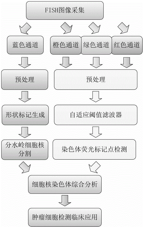
[0147] Using the FISH image parallel processing and analysis method based on the adaptive shape marker watershed algorithm of the present invention, the obtained FISH image is analyzed, such as figure 1 It mainly includes the following steps:

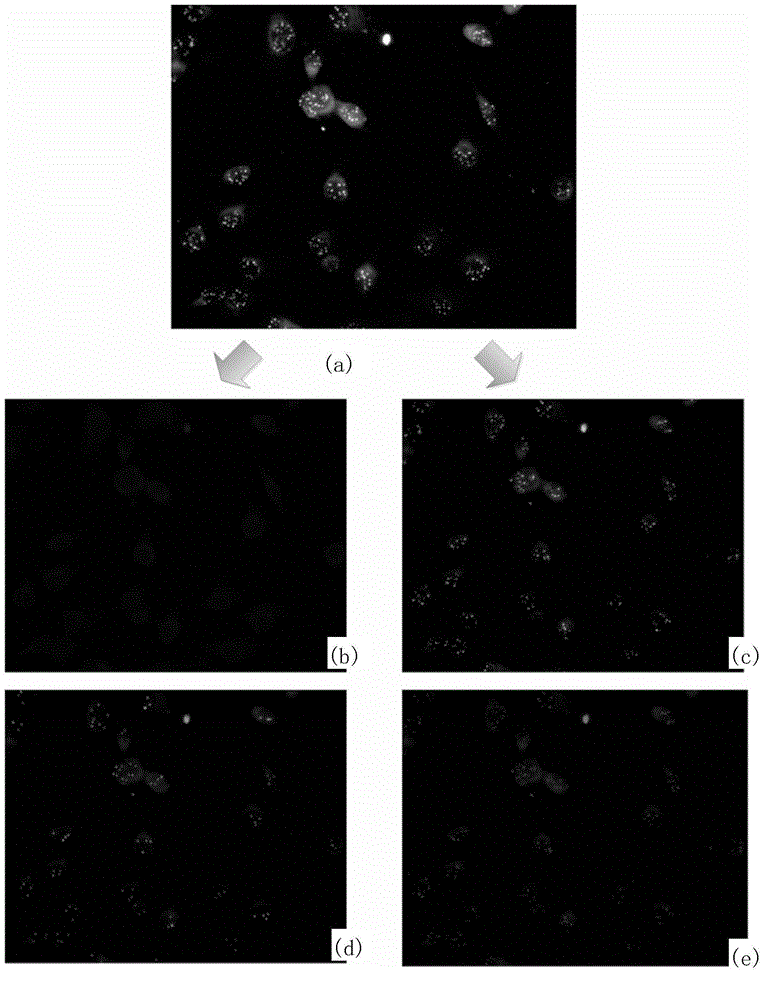
[0148] 1. Preprocess FISH images derived from tumor tissue sections, including image decomposition and grayscale, to obtain grayscale images containing only nuclei and grayscale images containing only orange, green, and red chromosomal fluorescent markers respectively. , the specific implementation process is divided into five steps as follows (see appendix figure 2 ):

[0149] 1.1 Read the original FISH color RGB image in the form of a three-dimensional matrix, where the third d...

Embodiment 2

[0175] Example 2 FSIH detection of circulating tumor cells

[0176] In Example 2, after whole blood cells were lysed red blood cells, a large number of leukocytes were removed by negative enrichment, nuclei were stained with DAPI, chromosomes were stained with orange-labeled probes, and leukocytes were stained with red-labeled antibodies. Circulating tumor cells were detected using substantially the same method as in Example 1. In Example 2, the circulating tumor cell images containing 60 cells were detected, and the processing speed was 5 seconds per image on a common desktop computer with 2.4GHz CPU and 4GB memory, and the detection accuracy of tumor cells was 92.9%, which was the same as the conventional method. Compared with this, the detection speed and accuracy are significantly improved.

[0177] The schematic diagram of the present invention for FISH detection of circulating tumor cells is as follows Figure 8 where (a) is the blue monochromatic image including the n...


PUM


Abstract

The invention provides a fluorescence in situ hybridization (FISH) image parallel processing and analysis method. The method employs parallel processing, detection of chromosome fluorescent labeling points and edge detection and segmentation of nucleuses can be performed simultaneously, and the processing time is reduced; and a watershed algorithm based on adaptive shape labels is employed so that the segmentation precision of the adhered nucleuses in FISH images is substantially improved, the detection accuracy of relative positions of the nucleuses in tumor cells and the chromosome fluorescent label points is improved, and online real-time detection of the tumor cells can be realized.

Description

technical field [0001] The invention belongs to the field of biomedicine, in particular to a parallel image processing and analysis method of fluorescence in situ hybridization (FISH). Background technique [0002] Fluorescence in situ hybridization is the process of in situ hybridization of several different colored fluorescein-labeled probes alone or in combination to detect several specific nucleic acid sequences in interphase cells or metaphase cells at the same time. FISH images collected by electron microscopy can simultaneously detect multiple genes, distinguish complex chromosomal translocations and small deletions, and provide a reliable detection tool for cytogenetic research. [0003] In cancer research, FISH technology is widely used for tumor cell detection in tumor tissue sections or circulating tumor cells (CTCs). In such biomedical applications, due to the huge number of cells contained in FISH images and the uneven distribution of nuclei, mutual adhesion of...

Claims


Application Information

Patent Timeline
IPC IPC(8): G06T7/00G06T7/40
CPCG06T7/0012G06T2207/20152G06T2207/30024G06T2207/30096
Inventor 时鹏张鲁榕洪金省
Owner XIAMEN LUJIA BIOTECH
Learn More