
Brain tumor image segmentation method based on multi-sequence MR (Magnetic Resonance) image correlation information

A technology of correlating information and image segmentation, applied in the field of biomedical image processing, it can solve the problems of boundary destruction, segmentation effect dependent on seed point selection and growth order, automatic segmentation of lung tumors, etc.

Inactive Publication Date: 2016-12-07
GUANGDONG POLYTECHNIC NORMAL UNIV
View PDF9 Cites 16 Cited by
  • Summary
  • Abstract
  • Description
  • Claims
  • Application Information

AI Technical Summary

Problems solved by technology

The inherent disadvantage of region growth is that the segmentation effect depends on the selection of seed points and the growth sequence. The disadvantage of region splitting technology is that the boundary may be destroyed
[0008] The patent "a lung tumor segmentation method based on graph cut PET and CT images, application number CN201410140351.2" mainly solves the problem of automatic lung tumor segmentation of existing PET and CT images

Method used


Image

  • Brain tumor image segmentation method based on multi-sequence MR (Magnetic Resonance) image correlation information
  • Brain tumor image segmentation method based on multi-sequence MR (Magnetic Resonance) image correlation information
  • Brain tumor image segmentation method based on multi-sequence MR (Magnetic Resonance) image correlation information

Examples


Embodiment Construction

[0035] The present invention will be described in more detail and complete below in conjunction with the accompanying drawings and specific embodiments. It should be understood that the specific embodiments described here are only used to explain the present invention, but not to limit the present invention.

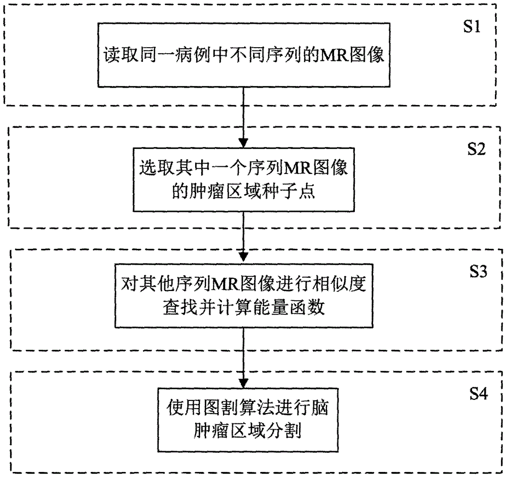
[0036] like figure 1 As shown, a kind of brain tumor image segmentation method based on multi-sequence MR image correlation information of the present invention comprises the following steps:

[0037] S1: Obtain MR images of different sequences in the same case, including T1 (longitudinal relaxation time), T2 (transverse relaxation time), T2WI+FLAIR (T2-weighted imaging), and T1WI (T1-weighted imaging) four imaging sequences.

[0038] S2: Select the brain tumor area of ​​one of the serial MR images as the tumor seed point. The seed point selection method only needs to select one of the serial MR images, and does not need to be selected in all the serial MR images. The s...


PUM


Abstract

The invention puts forward a brain tumor image segmentation method based on multi-sequence MR (Magnetic Resonance) image correlation information. The method comprises the following steps of: S1: reading the MR images of different sequences in the same focus; S2: selecting the brain tumor area of the MR image of one sequence as a tumor seed point; S3: carrying out similarity matching on the MR images of other sequences by the selected brain tumor seed point, and searching potential brain tumor seed points and background seed points in the MR images of other sequences; and S4: taking the searched potential brain tumor seed points as priori knowledge, and utilizing an image segmentation algorithm to realize the accurate segmentation of the brain tumor area in the multi-sequence MR image.

Description

technical field [0001] The invention relates to a brain tumor MR image segmentation method based on graph cut theory, and belongs to the field of biomedical image processing. Background technique [0002] MRI (Magnetic Resonance Imaging, MRI) is a type of tomographic imaging, which uses magnetic resonance phenomena to obtain electromagnetic signals from the human body and reconstruct human body information. MR images have excellent characteristics such as high contrast to soft tissue, direct layered imaging in any direction, non-invasive, non-invasive, and high spatial resolution. It has become the preferred computer-aided diagnostic method in the diagnosis of brain tumors and is widely used in medical diagnosis. , treatment, preoperative planning, postoperative monitoring and other important links. [0003] In the diagnosis of brain tumors based on MR images, it is usually necessary to refer to four imaging sequences of T1 (longitudinal relaxation time), T2 (transverse rel...

Claims


Application Information

Patent Timeline
IPC IPC(8): G06T7/00
CPCG06T2207/30016G06T2207/30096G06T2207/10088
Inventor 梁鹏郑振兴吴玉婷林智勇贾西平
Owner GUANGDONG POLYTECHNIC NORMAL UNIV
Learn More