Fluorescence probe for detecting pH of lysosome in cancer cells
A fluorescent probe and endolysosome technology, applied in the field of analytical chemistry, can solve the problems of lack of lysosome targeting, achieve good fluorescence emission spectrum characteristics, easy promotion, and broad application prospects
- Summary
- Abstract
- Description
- Claims
- Application Information
AI Technical Summary
Problems solved by technology
Method used
Image
Examples
Embodiment 1
[0029] Preparation of fluorescent probe for detecting lysosome pH in cancer cells according to the present invention
[0030] Compound a1 (1 mmol), a2 (1 mmol), 1-(3-dimethylaminopropyl)-3-ethylcarbodiimide (2 mmol), 1-hydroxybenzene Add triazole (1.5 mmol), triethylamine (2.2 mmol) and N,N-dimethylformamide (5 mL) into a 50 mL one-necked flask, stir at room temperature for 24 hours, filter, and dry. The obtained solid is purified by column chromatography to obtain a light yellow product, which is the ratiometric fluorescent probe for detecting intracellular lysosome pH according to the present invention. The resulting solid was purified by column chromatography (dichloromethane:methanol=20:1) to obtain a yellow product, which is the fluorescent probe 1a for detecting the pH of lysosomes in cancer cells according to the present invention, with a yield of 56%.
[0031] The above-mentioned fluorescent probes for detecting intracellular lysosome pH 1 H NMR spectrum see figure ...
Embodiment 2
[0033] The fluorescence spectrum of the fluorescent probe for detecting lysosome pH in cancer cells according to the present invention under different pH conditions
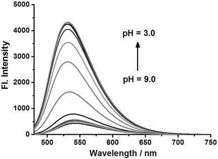
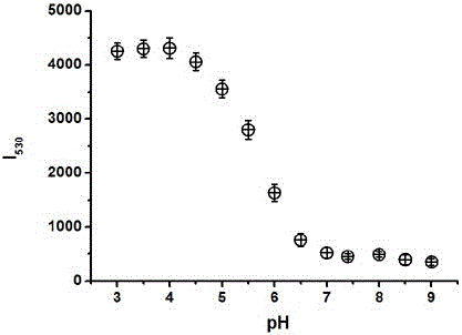
[0034] Prepare in advance 10 parts of 5 mL of 5 μM probe B-F buffer solution, containing 5% methanol, pH 3.0, 3.5, 4.0, 4.5, 5.0, 5.5, 6.0, 6.5, 7.0, 7.5, 8.0, 8.5, 9.0. Fluorescence detection is then performed (λ Ex = 445 nm); calculate the fluorescence intensity in each system; evaluate the response performance of the probe to pH (see figure 2 ). When the pH of the analysis solution rises from 3.0 to 9.0, the fluorescence intensity at 531nm is gradually weakened, see image 3 . Among them, when the pH of the analysis solution is in the range of 4.5~6.5, the linear relationship between the fluorescence intensity at 531nm and the pH can be expressed by the formula Y = 12022 - 1723 * X (R = 0.9886), where Y is the fluorescence intensity and X is the pH value.
Embodiment 3
[0036] Fluorescence spectra of fluorescent probes reacted with different substances
[0037] Prepare 10 parts of 5 mL of 5 μM probe B-F buffer solution (containing 5% methanol, pH = 7.4), and then add 50 μL of 10 mM Hcys, Na 2 S, Na 2 SO 3 , NO, H 2 o 2 , HClO 4 , Cys, GSH, NaNO 2 , VC, etc. in PBS solution. Fluorescence detection is then performed (λEx = 445 nm); calculate the fluorescence intensity in each system; evaluate the interference of the different substances on the fluorescent probe solution (see Figure 4 ). pass Figure 4 It can be seen that when the pH of the solution is 4.5 or 7.4, adding Hcys, Na 2 S, Na 2 SO 3 When small molecules are used, the fluorescence intensity of the solution remains basically unchanged, which means that the detection results are basically not affected by other interfering substances.
PUM
 Abstract
Description
Claims
Application Information
 - R&D Engineer
- R&D Manager
- IP Professional
- Industry Leading Data Capabilities
- Powerful AI technology
- Patent DNA Extraction
Browse by: Latest US Patents, China's latest patents, Technical Efficacy Thesaurus, Application Domain, Technology Topic, Popular Technical Reports.
© 2024 PatSnap. All rights reserved.Legal|Privacy policy|Modern Slavery Act Transparency Statement|Sitemap|About US| Contact US: help@patsnap.com


