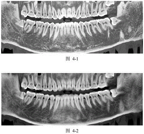
Method for extracting panoramic image from three-dimensional conical beam CT data of dentistry department
A cone-beam and panorama technology, which is used in the generation of X-ray panoramas and the field of panorama extraction from 3D dental cone-beam CT data, can solve the problems of reducing image quality, affecting diagnostic accuracy, and image blurring, reducing Manual operation, avoid image blur, improve the effect of precision
- Summary
- Abstract
- Description
- Claims
- Application Information
AI Technical Summary
Problems solved by technology
Method used
Image
Examples
Embodiment 1
[0052] A method for extracting panoramas from three-dimensional dental cone-beam CT data, the steps of which are as follows:
[0053] ⑴ Generate dental arch curves based on 3D dental cone beam CT data;
[0054] (2) Extract the three-dimensional oral panoramic surface according to the generated dental arch curve;
[0055] (3) Expand the three-dimensional oral panorama surface to obtain the oral panorama.
[0056] Preferably, the specific steps of generating the dental arch curve according to the three-dimensional dental cone-beam CT data in the step (1) are: calculating the maximum density projection image of the three-dimensional CT data along the vertical direction, separating the dental arch in the maximum density projection image, and Fit the arch curve.
[0057] Preferably, the specific step of separating the dental arch in the maximum density projection image is: separating the dental arch in the maximum density projection image by combining a grayscale-based segmentati...
Embodiment 2
[0064] A method for extracting a panorama from three-dimensional dental cone-beam CT data can extract a three-dimensional oral panorama curved surface, and expand the three-dimensional curved surface to obtain an oral panorama.
[0065] The method flow is as follows figure 1 As shown, the specific steps are as follows:
[0066] 1. Generate the dental arch curve according to the three-dimensional dental cone beam CT data. The specific implementation method is as follows:
[0067] Calculate the MIP image of the 3D CT data along the vertical direction, such as Figure 2-2 shown;
[0068] Apply the k-means++ clustering method on the maximum density projection image to extract the brightest bone area after clustering; calculate the center of gravity point of the bone area, and its quadrilateral neighborhood, so that the quadrilateral neighborhood covers p=75% of the bone area, Get the coordinate range represented by the quadrilateral neighborhood; apply the coordinate threshold ...
PUM
 Abstract
Description
Claims
Application Information
 - R&D
- Intellectual Property
- Life Sciences
- Materials
- Tech Scout
- Unparalleled Data Quality
- Higher Quality Content
- 60% Fewer Hallucinations
Browse by: Latest US Patents, China's latest patents, Technical Efficacy Thesaurus, Application Domain, Technology Topic, Popular Technical Reports.
© 2025 PatSnap. All rights reserved.Legal|Privacy policy|Modern Slavery Act Transparency Statement|Sitemap|About US| Contact US: help@patsnap.com


