
Method for evaluating exposure dosage of digital mammary gland X-ray imaging system

An imaging system and exposure dose technology, applied in the field of medical image processing, can solve problems such as the inability to evaluate the exposure dose, and achieve the effect of simple algorithm, convenient use, and improved accuracy

Active Publication Date: 2015-12-30
深圳蓝影医学科技股份有限公司
View PDF2 Cites 7 Cited by
  • Summary
  • Abstract
  • Description
  • Claims
  • Application Information

AI Technical Summary

Problems solved by technology

This method can automatically calculate the appropriate exposure parameters, but cannot evaluate the exposure dose

Method used


Image

  • Method for evaluating exposure dosage of digital mammary gland X-ray imaging system
  • Method for evaluating exposure dosage of digital mammary gland X-ray imaging system
  • Method for evaluating exposure dosage of digital mammary gland X-ray imaging system

Examples


Embodiment Construction

[0036] The present invention will be described in further detail below in conjunction with the accompanying drawings, but the present invention is not limited only to the following embodiments. A method for evaluating the exposure dose of a digital mammography system according to the present invention specifically comprises the following steps:

[0037] 1. Exposure index correction:

[0038] Because different flat-panel detectors have different responses to dose, it is necessary to calibrate the exposure index before using different types of flat-panel detectors.

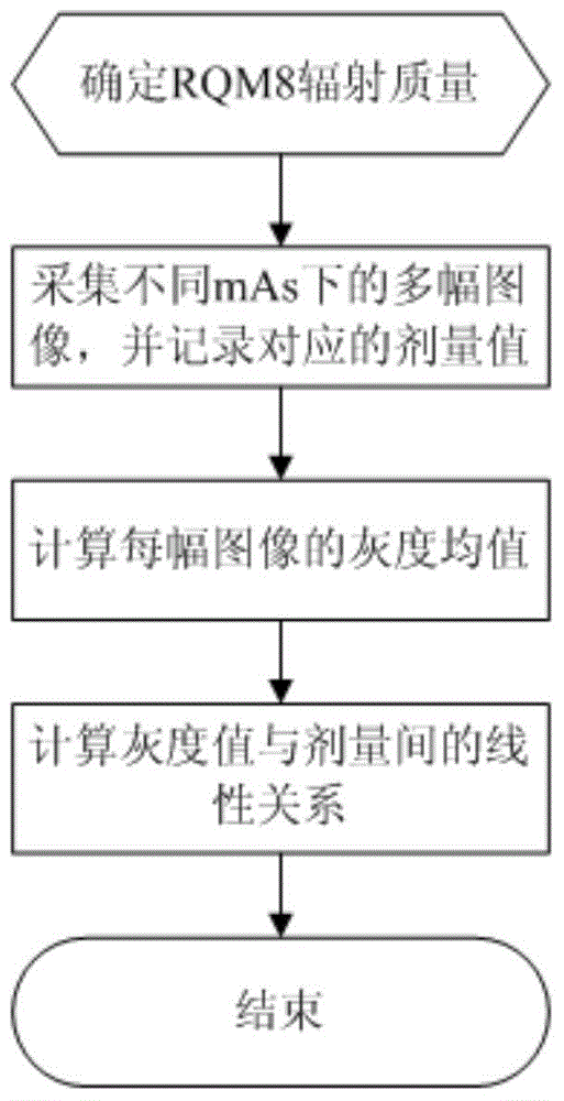
[0039] Place the dosimeter on the chest wall side of the flat panel detector, that is, the side close to the body of the patient when taking pictures, to determine the radiation quality of RQM8. RQM8 radiation quality is a related standard well known to researchers in the field, and will not be described here.

[0040] Images of 6 different mAs were collected, and the dose corresponding to each image was recorded....


PUM


Abstract

The invention discloses a method for evaluating the exposure dosage of a digital mammary gland X-ray imaging system. The method includes the following steps of setting out a target exposure index and calculating a deviation index DI. By means of the technical scheme, according to the deviation between the exposure index and a target exposure index, the exposure dosage of a mammary gland image is evaluated, and therefore a doctor can conveniently and subsequently adjust exposure parameters, an image of appropriate exposure dosage is obtained, the algorithm is simple, and use is convenient.

Description

technical field [0001] The invention relates to a medical image processing technology, in particular to an evaluation method for medical image exposure dose. Background technique [0002] As an important topic of women's health, mammary gland has been paid more and more attention by people. In recent years, the incidence of breast cancer has been increasing year by year in our country. In many big cities, breast cancer has become the most common malignant tumor in women, which seriously threatens women's health. Studies have shown that if breast cancer can be detected early, it will greatly improve Its cure rate, so the early diagnosis of breast cancer has very important clinical value. [0003] The actual clinical discovery of breast cancer early shows that there are tiny calcifications in the breast. These tiny calcifications are difficult to find when using ultrasound imaging, and the use of digital mammography to examine the breast is an effective method for early detec...

Claims


Application Information

Patent Timeline
Patent Type & Authority Applications(China)
IPC IPC(8): A61B6/00
Inventor 郭朋秦启兴鄢照龙李勇赵明
Owner 深圳蓝影医学科技股份有限公司
Learn More