Colloidal gold immunochromatography test paper for antibodies of pseudorabies viruses of gE, gB and gD and preparation method
A porcine pseudorabies virus and immunochromatographic detection technology, which is applied in biological testing, measuring devices, analytical materials, etc., to achieve the effects of simple operation, good detection repeatability, and easy portability
- Summary
- Abstract
- Description
- Claims
- Application Information
AI Technical Summary
Problems solved by technology
Method used
Image
Examples
Embodiment 1
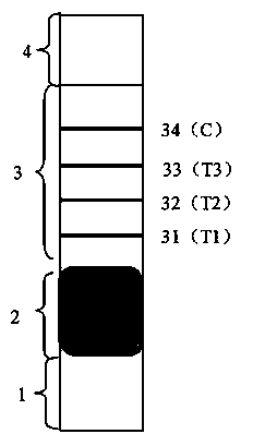
[0033] Colloidal gold immunochromatography test paper planar structure area map of porcine pseudorabies virus gE, gB and gD antibodies figure 1As shown, the horizontal plane of the test paper is as follows from bottom to top: sample absorption area 1, gold label probe area 2, immobilized antigen and antibody area 3 and water absorption area 4, and immobilized antigen and antibody area 3 is coated with detection lines T1 31, test line T2 32, test line T3 33 and control line C 34.
[0034] Colloidal gold immunochromatographic test paper longitudinal section structure diagram of porcine pseudorabies virus gE, gB and gD antibodies (specific materials are used to represent each area) as shown in figure 2 As shown, the polyethylene plate 5 with a layer of polyvinyl chloride lining film is the support plate, the glass fiber membrane 6 is the sample absorption area, the polyester film 7 is the gold standard probe area, and the nitrocellulose membrane 8 is the immobilized antigen and...
Embodiment 2
[0053] Except that the coating amount of the detection line T1 is 5 μg; the coating amount of the detection line T2 is 5 μg, the coating amount of the detection line T3 is 5 μg, the coating amount of the control line C is 10 μg, and the gold standard probe colloidal gold-labeled mouse anti-pig IgG single The amount of anti-labeling is 20 μg of monoclonal antibody labeled with colloidal gold per mL, and the coating volume of gold-labeled probe is 5 μL; the method of colloidal gold-labeled porcine circovirus type 2 monoclonal antibody: take colloidal gold with a radius of 10nm and a concentration of 0.01% respectively 20mL and mouse anti-pig IgG monoclonal antibody 400μg, combined by magnetic stirring and shaking under the condition of pH 9.0, adding bovine serum albumin (BSA) and polyethylene glycol 20000 (PEG20000) as stabilizers, and making BSA The final mass concentration was 2%, and the final mass concentration of PEG20000 was 0.05%. The unbound mouse anti-pig IgG monoclonal...
Embodiment 3
[0055] Except that the coating amount of the detection line T1 is 2 μg; the coating amount of the detection line T2 is 2 μg, the coating amount of the detection line T3 is 2 μg, the coating amount of the control line C is 5 μg, and the gold standard probe colloidal gold-labeled mouse anti-pig IgG single The amount of anti-labeling is 10 μg of monoclonal antibody labeled with colloidal gold per mL, and the coating volume of gold-labeled probe is 8 μL; the method of colloidal gold-labeled porcine circovirus type 2 monoclonal antibody: take colloidal gold with a radius of 20nm and a concentration of 0.01% respectively 20mL and mouse anti-pig IgG monoclonal antibody 200μg, combined by magnetic stirring and shaking under the condition of pH 9.0, adding bovine serum albumin (BSA) and polyethylene glycol 20000 (PEG20000) as stabilizers, and making BSA The final mass concentration was 2%, and the final mass concentration of PEG20000 was 0.05%. The unbound mouse anti-pig IgG monoclonal ...
PUM
 Abstract
Description
Claims
Application Information
 - Generate Ideas
- Intellectual Property
- Life Sciences
- Materials
- Tech Scout
- Unparalleled Data Quality
- Higher Quality Content
- 60% Fewer Hallucinations
Browse by: Latest US Patents, China's latest patents, Technical Efficacy Thesaurus, Application Domain, Technology Topic, Popular Technical Reports.
© 2025 PatSnap. All rights reserved.Legal|Privacy policy|Modern Slavery Act Transparency Statement|Sitemap|About US| Contact US: help@patsnap.com


