
Visual preparation method of polymeric fibers based on laser scanning confocal microscope

A polymer fiber, laser confocal technology, applied in the field of laser confocal microscope imaging, can solve problems such as inability to observe

Inactive Publication Date: 2013-06-26
SHANGHAI NAT ENG RES CENT FORNANOTECH
View PDF11 Cites 5 Cited by
  • Summary
  • Abstract
  • Description
  • Claims
  • Application Information

AI Technical Summary

Problems solved by technology

This results in thicker polymer fibers and other biomaterial matrices that cannot be observed
[0007] (2) Self-assembly of small molecules leads to indirect imaging of polymer fibers
However, its original intention is not to visualize polymer fibers for laser confocal microscope observation, but to judge the self-assembly ability of small molecules; at the same time, polymer fibers interact indirectly with cells through small molecules, not directly with polymer fibers. Cells act directly, which is not conducive to directly determine the influence of polymer fibers on the number, distribution and shape of cells

Method used


Image

  • Visual preparation method of polymeric fibers based on laser scanning confocal microscope
  • Visual preparation method of polymeric fibers based on laser scanning confocal microscope
  • Visual preparation method of polymeric fibers based on laser scanning confocal microscope

Examples


Embodiment 1

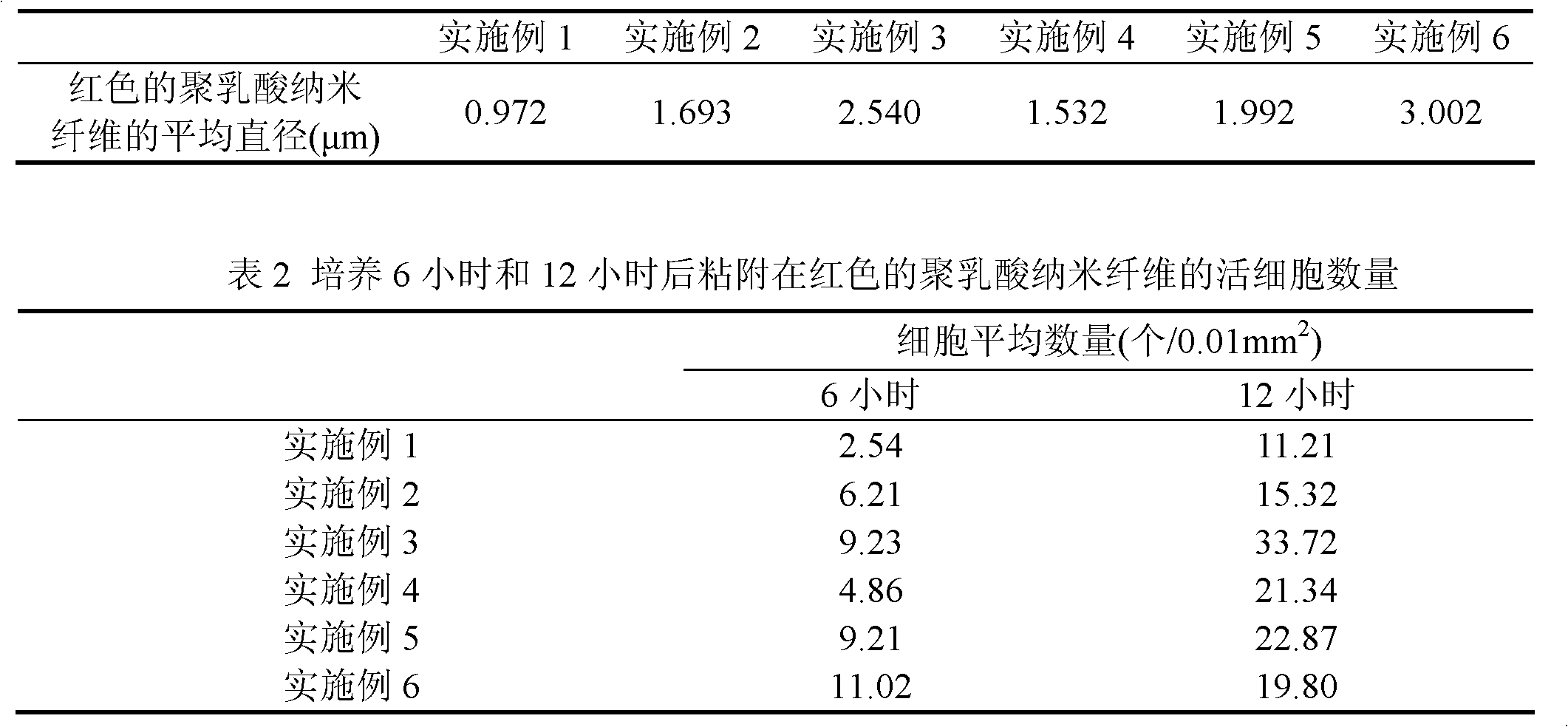
[0036] Weigh 0.2g of polylactic acid, put it into 20ml of dichloromethane solvent, heat and condense to reflux and dissolve for 1 hour until the copolymer is completely dissolved and the solution is colorless and transparent. Add 10 mg of red dye isothiocyanerhodamine B to the above solution, stir and dissolve until it becomes a red transparent liquid. Put it into the glass syringe in the spray device so that the injection rate is 0.3ml / hour, a high voltage of 10,000 volts is generated between the positive electrode of the needle tip and the negative electrode of the substrate, and the distance between the needle tip and the receiving device is 30mm, and a glass piece is used to collect the The polylactic acid fibers coated with isothiocyanerhodamine B ejected by electrospinning were collected for 15 minutes. Soak the polylactic acid nanofibers coated with isothiocyanato-rhodamine B in 200ml of pure water, and shake on a constant temperature shaker at room temperature for 3 da...

Embodiment 2

[0040] Weigh 1g of polylactic acid, put it into a mixed solvent of 18ml of dichloromethane and 2ml of N,N-dimethylformamide, heat and condense to reflux for 1 hour until the copolymer is completely dissolved and the solution is colorless and transparent. Add 20 mg of red dye isothiocyanerhodamine B to the above solution, stir and dissolve until it becomes a red transparent liquid. Put it into the glass syringe in the spraying device so that the injection rate is 0.3ml / hour, a high voltage of 5000 volts is generated between the positive electrode of the needle tip and the negative electrode of the substrate, and the distance between the needle tip and the receiving device is 20 mm, and the passing through is collected with a glass sheet. The polylactic acid fibers coated with isothiocyanerhodamine B ejected by electrospinning were collected for 120 minutes. Soak the polylactic acid nanofibers coated with isothiocyanato-rhodamine B in 400ml of pure water, and shake on a constant...

Embodiment 3

[0044] Weigh 1g of polylactic acid and 1g of polycaprolactone, put them into 20ml of dichloromethane solvent, heat and condense to reflux for 1 hour until the copolymer is completely dissolved and the solution is colorless and transparent. Add 100 mg of red dye isothiocyanerhodamine B to the above solution, stir and dissolve until it becomes a red transparent liquid. Put it into the glass syringe in the spraying device so that the injection rate is 0.1ml / hour, a high voltage of 5000 volts is generated between the positive electrode of the needle tip and the negative electrode of the substrate, and the distance between the needle tip and the receiving device is 15mm, and a glass piece is used to collect the The polylactic acid fibers coated with isothiocyanerhodamine B ejected by electrospinning were collected for 30 minutes. Soak the polylactic acid nanofibers coated with isothiocyanato-rhodamine B in 200ml of pure water, and shake on a constant temperature shaker at room temp...


PUM

PropertyMeasurementUnit
The average diameteraaaaaaaaaa

Abstract

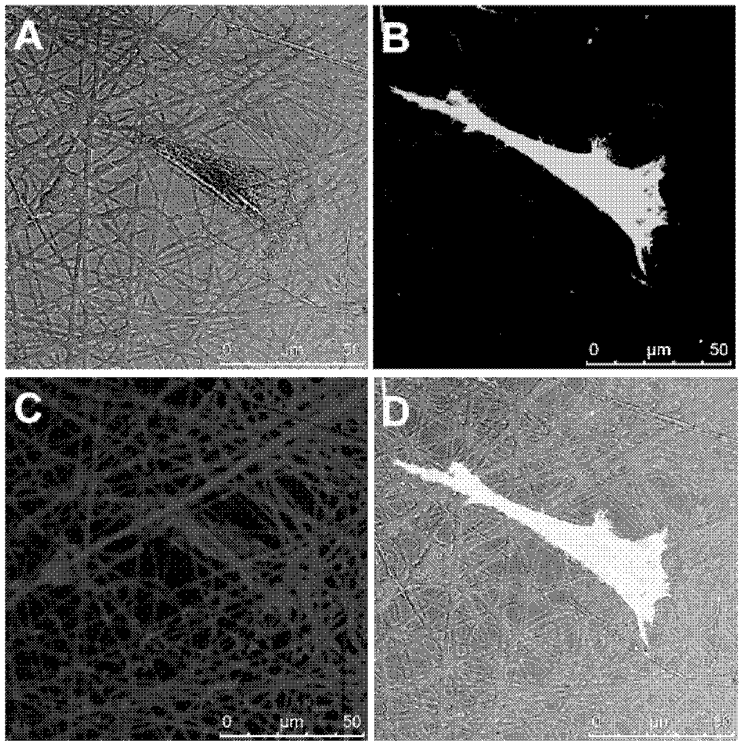
The invention relates to a visual preparation method of polymeric fibers based on a laser scanning confocal microscope. The visual preparation method is characterized by comprising the following steps of: dissolving a high polymer material into an organic solvent; adding a fluorescent dye to prepare an electrostatic spinning, thus obtaining the polymeric fibers wrapped with the fluorescent dye; dipping the prepared polymeric fibers into the water to wash to remove the fluorescent dye residual on the surface; carrying out ultraviolet sterilization; inoculating cells; marking the cells by the fluorescent dye; and observing by a laser confocal microscope. Compared with the simple cell laser confocal image, the laser confocal image prepared by the preparation method provided by the invention has the advantage that the polymeric fibers provide the support, thus the sense of space of the image is improved, and the image is much more real. By adopting the preparation method, the amount, distribution and forms of living cells on the polymeric fibers can be directly observed at the same time by utilizing the laser scanning confocal microscope; and a living cell culture chamber in match with the laser scanning confocal microscope also can be utilized, which enables the continuous and direct observation on the amount, distribution and forms of the living cells on the polymeric fibers to become possible.

Description

technical field [0001] The present invention relates to a preparation method for the visualization of polymer fibers, in particular to a preparation method for the visualization of polymer fibers under a laser confocal scanning microscope, which is used to directly observe the interaction between cells and polymer fibers, and belongs to the field of laser confocal microscope imaging . Background technique [0002] Laser confocal microscope is based on the imaging of fluorescence microscope with laser scanning device installed, using computer for image processing, and the resolution is increased by 30-40% on the basis of optical imaging, which expands the application range of traditional fluorescence microscope and becomes A new generation of powerful research tools in the fields of morphology, molecular biology, neuroscience, pharmacology, genetics and more. [0003] So far, in the field of tissue engineering, laser confocal microscopy is mainly used to observe: (1) the num...

Claims


Application Information

Patent Timeline
IPC IPC(8): G01N1/30
Inventor 魏岱旭钟建闫志强何丹农
Owner SHANGHAI NAT ENG RES CENT FORNANOTECH
Learn More