Marker for cancer detection and application thereof
A cancer detection and marker technology, applied in the field of molecular identification of cancer detection, can solve the problem of lack of molecular markers in cancer, and achieve the effect of high accuracy and high reference value
- Summary
- Abstract
- Description
- Claims
- Application Information
AI Technical Summary
Problems solved by technology
Method used
Image

Examples
Embodiment 1
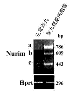
[0044] Example 1 RT-PCR detection of testicular seminoma
[0045] 1. Total RNA extraction
[0046] 1) Immediately take 50 mg of fresh tumor tissue, place it in a mortar, carefully pour an appropriate amount of liquid nitrogen, and grind it quickly until the tissue becomes white powder.
[0047] 2) Add 1 mL of Trizol (purchased from Invitrogen), mix well, and lyse at room temperature for 5 minutes.
[0048] 3) Add 0.2mL chloroform, mix well, centrifuge at 12000rpm for 15 minutes, take the upper aqueous phase into a new centrifuge tube.
[0049] 4) Add 0.5mL isopropanol, mix well, centrifuge at 12000rpm for 15 minutes, discard the supernatant, the precipitate is RNA.
[0050] 5) Wash with freshly prepared 75% ethanol, centrifuge at 12,000 rpm for 15 minutes, and discard the supernatant.
[0051] 6) Add 20-25 μL of deionized water, and measure the OD to calculate the RNA concentration after the precipitate is dissolved.
[0052] 2. Preparation of cDNA template. See Table 1 f...
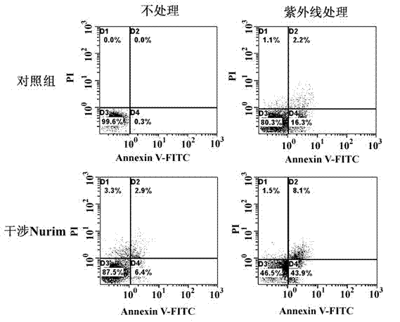
Embodiment 2
[0066] Example 2 Detection of various types of tumors by Western blot
[0067] 1. Extraction of protein
[0068] Soak the whole set of homogenizer in acid for more than 4 hours, wash and dry, pack with tin foil, and bake at 180°C for 2 hours. Take 100 mg of fresh tissue, add PBS, grind the tissue with a homogenizer, break the cells with an ultrasonic instrument, add the lysate, boil for 3 minutes, place on ice for 5 minutes, centrifuge at 5000rpm for 5 minutes, and take the supernatant for loading. Lysis buffer configuration, 50mmol / L Tris Base, 100mmol / L dithiothreitol, 2% (m / v) SDS, 0.1% bromophenol blue, 10% (v / v) glycerol.
[0069] 2. Western blot
[0070] 1) In the experiment, we used the SDS-PAGE gel separation system with a concentration of 15%, and the preparation methods are shown in Table 4 and Table 5. Electrophoresis buffer configuration, 25mmol / L Tris Base, 250mmol / L glutamic acid (pH8.3), 0.1% (m / v) SDS.
[0071] components Volume (mL) water ...
Embodiment 3
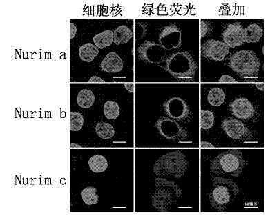
[0079] Example 3 Immunofluorescence detection of squamous cell carcinoma of the lung
[0080] 1. Frozen section
[0081] Pre-cool the cryostat to -20°C to -18°C. Choose the most suitable temperature according to the nature of different tissues. Serial sections were made with a thickness of 7 microns.
[0082] 2. Immunofluorescence
[0083] 1) Place frozen sections in pre-cooled methanol and fix at 4°C for 20 minutes. After fixation, wash three times with PBS at room temperature, 10 minutes each time. PBS configuration, 8 g NaCl, 0.2 g KCl, 1.44 g Na 2 HPO 4 , 0.24 g KH 2 PO 4 , adjust the pH to 7.4 with HCl, add water to 1 liter, and autoclave for 20 minutes.
[0084] 2) Put in 0.5%v / v TX-100, penetrate at room temperature for 20 minutes. Wash three times with PBS, 10 minutes each time.
[0085] 3) Add normal goat serum blocking solution and block for 1 hour at room temperature.
[0086] 4) join Nurim Primary antibody (generally the concentration is 1:100 dissolve...
PUM
| Property | Measurement | Unit |
|---|---|---|
| Thickness | aaaaa | aaaaa |
Abstract
Description
Claims
Application Information
 - R&D
- Intellectual Property
- Life Sciences
- Materials
- Tech Scout
- Unparalleled Data Quality
- Higher Quality Content
- 60% Fewer Hallucinations
Browse by: Latest US Patents, China's latest patents, Technical Efficacy Thesaurus, Application Domain, Technology Topic, Popular Technical Reports.
© 2025 PatSnap. All rights reserved.Legal|Privacy policy|Modern Slavery Act Transparency Statement|Sitemap|About US| Contact US: help@patsnap.com