Extraction and multiplication culture method and application of urine mesenchymal stem cells
A technique for the expansion and cultivation of stem cells, which is applied in the fields of cell biology and regenerative medicine, can solve the problems of high cost, restrictions on wide application, invasive separation and extraction methods, etc., achieve good monitoring or tracking, and the method is simple, easy, and good The effect of amplifying capacity
- Summary
- Abstract
- Description
- Claims
- Application Information
AI Technical Summary
Problems solved by technology
Method used
Image
Examples
Embodiment 1
[0042] Isolation and extraction of urinary mesenchymal stem cells from clean midstream urine:
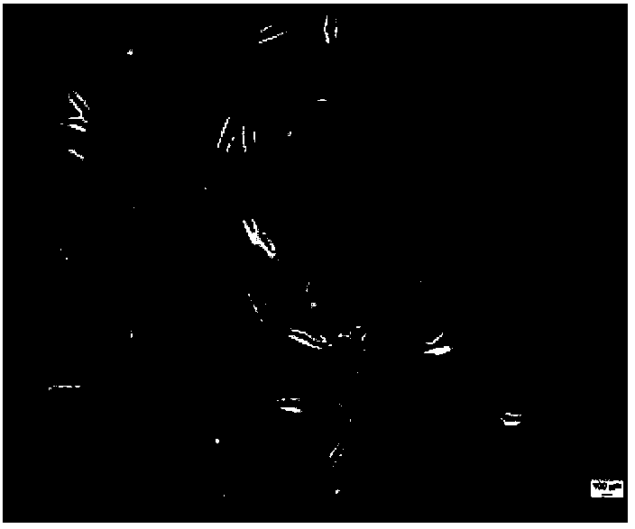
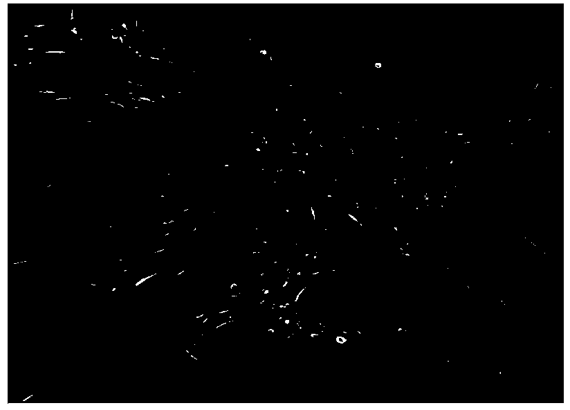
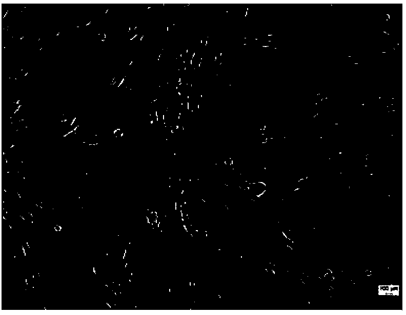
[0043] Under sterile conditions, take 200-250ml of clean middle urine from healthy volunteers. To prevent infection, add 5ml of double antibody containing penicillin 100U / ml + streptomycin 0.1mg / ml, and separate and extract immediately. Divide the clean urine into 50ml centrifuge tubes, centrifuge at 400g for 10min, and discard the supernatant. Wash twice by centrifugation at 400 g for 10 min with PBS. Take 100 μL of cell resuspension, use trypan blue to detect cell viability and count, adjust cell density, inoculate into a six-well plate containing serum-free medium, and place at 37°C in 5% CO 2 cultured in a humidified incubator.
[0044] After 3-7 days of culture, observe under a phase-contrast microscope. If bacterial contamination is observed, discard it. If there is no contamination, continue the culture. A single cell can be found to adhere to the wall, and the position of ...
Embodiment 2
[0046] Differentiation of urinary mesenchymal stem cells into osteocytes:
[0047] (1) Take the fourth-generation culture of urine mesenchymal stem cells obtained in Example 1
[0048] (2) Induction culture medium containing 0.1 μmol / L of dexamethasone, 10 nmol / L of vitamin D, and 10 mmol / L of β-glycerol phosphate was added to the urinary mesenchymal stem cells for induction culture, and the medium was changed twice a week.
[0049] (3) Alizarin red staining was performed to observe calcium nodules after 21 days of induced differentiation, and RT-PCR was used to detect the expression of osteopontin, osteocalcin, and bone morphogenetic protein.
[0050] The results showed that a large number of calcium nodules appeared in the induced cells (see Figure 12 ), the expression levels of osteopontin, osteocalcin, bone morphogenetic protein and other genes were significantly increased, indicating that urinary mesenchymal stem cells can differentiate into osteoblasts.
Embodiment 3
[0052] Differentiation of urinary mesenchymal stem cells into chondrocytes:
[0053] (1) Take the fourth-generation culture of urine mesenchymal stem cells obtained in Example 1
[0054] (2) Add induction medium containing transforming growth factor β 110 ng / ml, insulin-like growth factor 100 ng / ml and dexamethasone 0.1 μmol / L to urinary mesenchymal stem cells
[0055] (3) After induction and culture for 28 days, the extracellular matrix of the cells was stained with Alcian blue, and the expression of type II collagen was detected by immunohistochemistry.
[0056] The results showed that most of the induced and cultured urinary mesenchymal stem cells were stained with Alcian blue, and the expression of type II collagen was enhanced, indicating that the urinary mesenchymal stem cells could differentiate into chondrocytes.
PUM
 Abstract
Description
Claims
Application Information
 - R&D
- Intellectual Property
- Life Sciences
- Materials
- Tech Scout
- Unparalleled Data Quality
- Higher Quality Content
- 60% Fewer Hallucinations
Browse by: Latest US Patents, China's latest patents, Technical Efficacy Thesaurus, Application Domain, Technology Topic, Popular Technical Reports.
© 2025 PatSnap. All rights reserved.Legal|Privacy policy|Modern Slavery Act Transparency Statement|Sitemap|About US| Contact US: help@patsnap.com


