Method for detecting mite allergen specific antibody in blood serum
An allergen-specific technology, applied in the field of diagnosis, can solve the problems of low sensitivity and accuracy, and achieve the effects of improving sensitivity, improving detection efficiency, and shortening detection time
- Summary
- Abstract
- Description
- Claims
- Application Information
AI Technical Summary
Problems solved by technology
Method used
Image

Examples
Embodiment 1
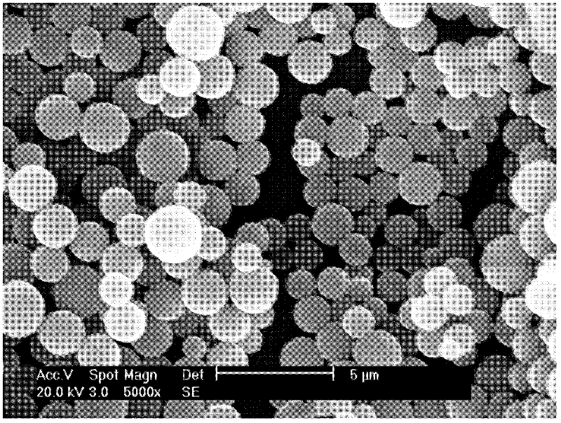
[0019] Sodium oleate-coated Fe was prepared using a conventional chemical co-precipitation method 3 o 4 Magnetic fluid: prepare 0.12mol / L ferrous chloride (FeCl 2 .4H 2 O) and 0.2mol / L ferric chloride (FeCl 3 .6H 2 O) 25 mL each, transferred to a 250 mL three-neck flask, stirred under nitrogen protection, and the stirring speed was 500 rpm. After raising the temperature of the above mixture to 55°C for 15 minutes, quickly add 3mol / L NaOH to adjust the pH value to 10-12. The temperature of the mixture was raised to 65° C., and the stirring was maintained for 40 min. Then slowly add 4% sodium oleate 50mL, raise the temperature to 90°C, continue to stir for 30min, and cool to room temperature to obtain Fe coated with sodium oleate. 3 o 4 ferrofluid. Fe of the ferrofluid 3 o 4 The particles are superparamagnetic, with an average particle size of 30-50nm.
[0020] Preparation of carboxyl-modified magnetic microspheres by dispersion polymerization: take 4mL of the above-m...
Embodiment 2
[0022] Coupling mite allergen protein to prepare immunomagnetic microspheres.
[0023] (1) Using 25mM MES (pH=6) as a buffer solution, wash the magnetic microspheres (with a concentration of 10mg / mL) twice. Dissolve the mite allergen protein with the above-mentioned buffer solution;
[0024] (2) Use freshly prepared EDC and NHS as the cross-linking agent, the concentration of EDC and NHS is 50 mg / mL, add a certain volume of cross-linking agent to the washed magnetic microspheres, and add 20 μL of cross-linking agent to each mg of magnetic microspheres. Combined agent, incubated with tilt rotation at room temperature for 30min to activate the magnetic microspheres;
[0025] (3) The activated magnetic microspheres were washed twice with MES buffer, then an appropriate amount of mite allergen protein was added (the mass ratio of magnetic microspheres to mite allergen protein was 15:1), vortexed and incubated at room temperature for 1 -2h;
[0026] (4) Wash the magnetic microsp...
Embodiment 3
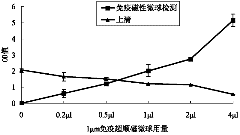
[0030] (1) Cleaning and activation of immunomagnetic microspheres: Take the immunomagnetic microspheres prepared and preserved above in a clean centrifuge tube, add PBS activation buffer containing 0.1% BSA in a volume ratio of 1:10, and mix well. Magnetically separate and remove buffer. Wash once more in this way, and then add the above-mentioned activation buffer to make up to the original demand.
[0031] (2) Sample incubation: Take 0.2 μL, 0.5 μL, 1 μL, 2 μL, and 4 μL of the above-mentioned cleaned and activated immunomagnetic microspheres (10 mg / mL), mix each with 100 μL of standard mite serum samples in each well of the microplate, and gently Shake gently and incubate with shaking for 15-20 minutes at room temperature.
[0032](3) Plate washing: After the incubation, magnetically separate, keep the supernatant in a new centrifuge tube for detection, add 200 μL of PBST buffer to each well to wash the plate, remove the supernatant after magnetic separation, and wash 3 tim...
PUM
| Property | Measurement | Unit |
|---|---|---|
| particle size | aaaaa | aaaaa |
| particle diameter | aaaaa | aaaaa |
| particle diameter | aaaaa | aaaaa |
Abstract
Description
Claims
Application Information
 - R&D
- Intellectual Property
- Life Sciences
- Materials
- Tech Scout
- Unparalleled Data Quality
- Higher Quality Content
- 60% Fewer Hallucinations
Browse by: Latest US Patents, China's latest patents, Technical Efficacy Thesaurus, Application Domain, Technology Topic, Popular Technical Reports.
© 2025 PatSnap. All rights reserved.Legal|Privacy policy|Modern Slavery Act Transparency Statement|Sitemap|About US| Contact US: help@patsnap.com