Method for displaying animal (bovine) heart conducting system
A technology of conduction system and heart, which is applied in the field of casting to display the three-dimensional structure of animal heart conduction system, which can solve the problems of inability to inject fillers, etc., and achieve the effect of facilitating the walking position and distribution of the general structure, easy to operate, and not easy to fade
- Summary
- Abstract
- Description
- Claims
- Application Information
AI Technical Summary
Problems solved by technology
Method used
Image

Examples
Embodiment 1
[0015] [Example 1] Dissection method
[0016] 1. Preparation of general materials Fresh beef heart, 5-10% liquid green plastic filler (recipe: solute: perchlorethylene 5-10g; solvent: cyclohexanone 100ml; shrinkage agent: dibutyl phthalate 3 ~5ml; 0.5~1.0g of Shanghai Marley Chlorine Oil Paint (Model: E1387), add the solute into the solvent, let it stand for 2 days to fully dissolve, then add shrinkage agent and Chlorine Oil Paint, and stir while adding , fully dissolved before use), 5 ~ 10ml syringes and needles, high-quality carbon ink, general scalpel instruments, etc.
[0017] 2. The right ventricle is cut from the anterior wall of the pulmonary artery to the conus pulmonary artery, then cut parallel to the right along the right coronary artery to the right heart border, and then cut along the right heart border to the apex of the heart. For the left ventricle, enter the knife from the midline of the anterior and posterior papillary muscles, cut to the bottom and apex of ...
Embodiment 2
[0018] [Example 2] perfusion method
[0019] 1. Take out the beef heart that has been refrigerated for 24 hours, and rinse it under running water for 80-100 minutes.
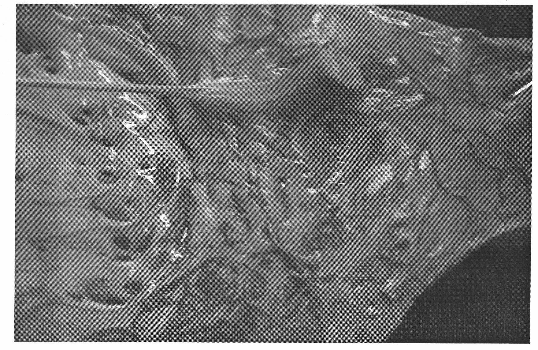
[0020] 2. Carefully look for the grayish-white or slightly transparent filament structure under the endocardium, or identify the Purkinje fibers after foaming the endocardium with an iodine cotton swab.
[0021] 3. Hold the syringe fully filled with the prepared plastic filler, and press the needle tip of the syringe parallel to the endocardium with the filamentous structure with a little force, so that the endocardium is slightly depressed, while keeping the slope of the needle tip upward. Advance the needle so that the bevel of the needle tip completely enters the subendocardium, and then advance the needle 1 to 2 mm. Turn the syringe 180 smoothly so that the bevel of the needle tip is directed toward the myocardium.
[0022] 4. Then try to push the piston lightly, and the filler can enter by pushing lightly...
Embodiment 3
[0024] [Example 3] Corrosion flushing
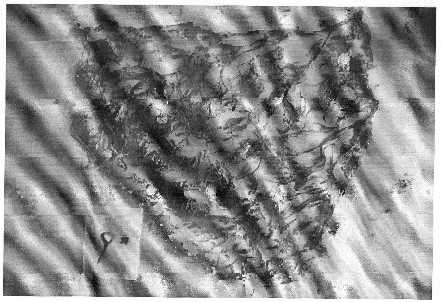
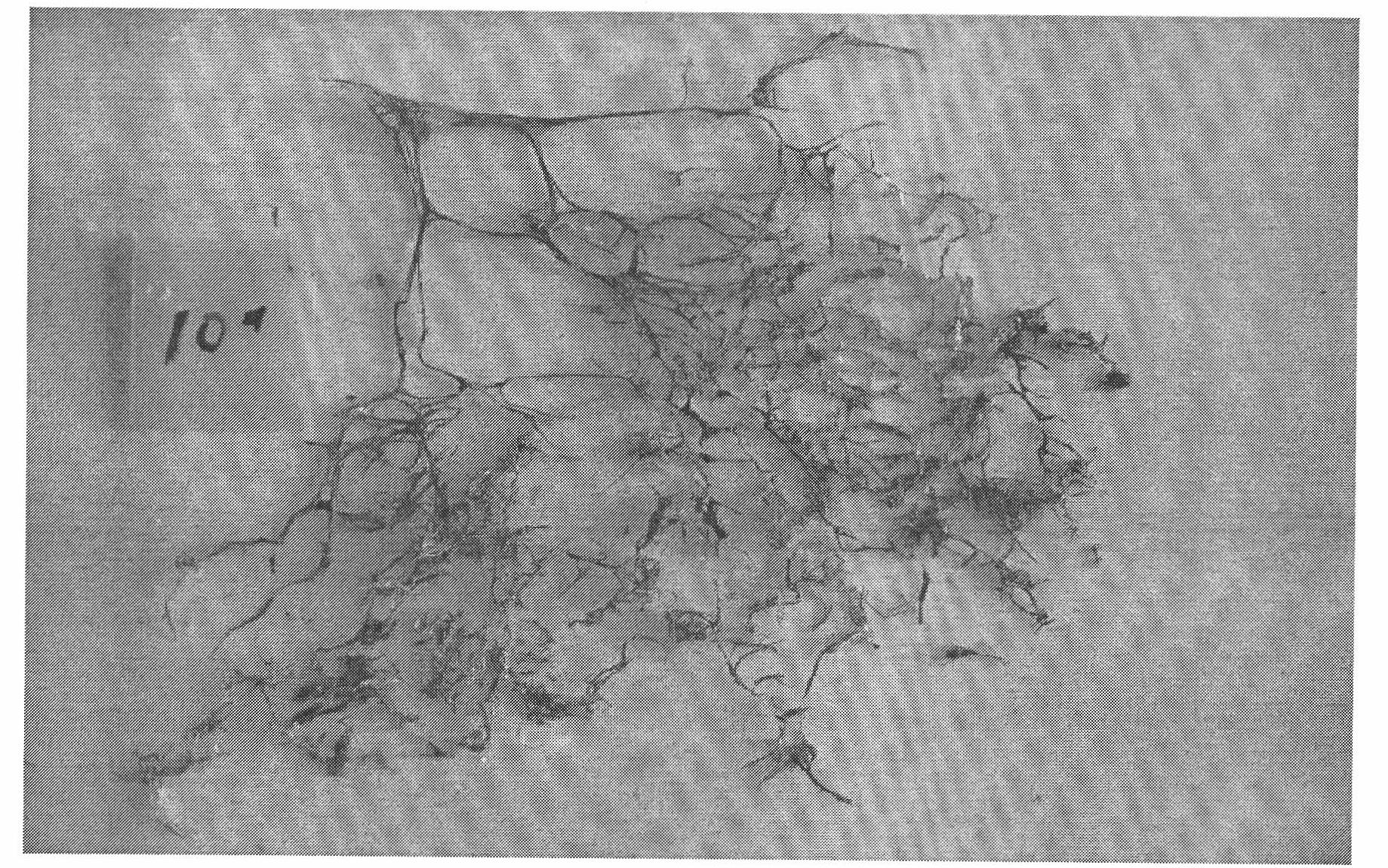
[0025] The perfused heart was allowed to stand naturally for 48 hours, and then it was moved to 50% hydrochloric acid solution for corrosion. After 72 hours, take it out and wash it with running water to wash away the decayed cardiac tissue, and keep the blue plastic cast bracket of the cardiac conduction system (left and right bundle branches and Purkinje fibers).
PUM
 Abstract
Description
Claims
Application Information
 - R&D
- Intellectual Property
- Life Sciences
- Materials
- Tech Scout
- Unparalleled Data Quality
- Higher Quality Content
- 60% Fewer Hallucinations
Browse by: Latest US Patents, China's latest patents, Technical Efficacy Thesaurus, Application Domain, Technology Topic, Popular Technical Reports.
© 2025 PatSnap. All rights reserved.Legal|Privacy policy|Modern Slavery Act Transparency Statement|Sitemap|About US| Contact US: help@patsnap.com