Eureka AIR delivers breakthrough ideas for toughest innovation challenges, trusted by R&D personnel around the world.

Method for separating heart stem cells from brown fat and splitting cardioblast

A technology of cardiac stem cells and brown fat, applied in animal cells, vertebrate cells, bone/connective tissue cells, etc., can solve the problems of no reports, etc., and achieve the effect of increasing yield, high yield, and strong implementability

Active Publication Date: 2010-05-12
INST OF BASIC MEDICAL SCI ACAD OF MILITARY MEDICAL SCI OF PLA
View PDF0 Cites 8 Cited by
  • Summary
  • Abstract
  • Description
  • Claims
  • Application Information

AI Technical Summary

Problems solved by technology

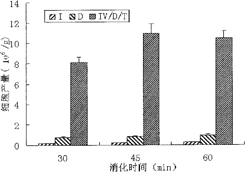
But the challenge is to find a suitable combination of digestive enzymes and appropriate digestion conditions, and can obtain the desired cell type and yield of viable cells (Pilgaard L et al., "Comparative analysis of highly defined proteases for the isolation of adipose tissue-derived stem cells", Regen Med., 2008, 3(5): 705-15)
However, such an ideal separation method has not been reported so far.

Method used


Image

  • Method for separating heart stem cells from brown fat and splitting cardioblast
  • Method for separating heart stem cells from brown fat and splitting cardioblast
  • Method for separating heart stem cells from brown fat and splitting cardioblast

Examples


Embodiment 1

[0038] Example 1 The method for isolating cardiac stem cells using rat brown adipose tissue as a tissue sample

[0039] (1) Rat brown adipose tissue collection and processing

[0040] Juvenile SD rats at 1-2 weeks after birth were sacrificed by cervical dislocation. Remove the hair near the scapula and sterilize by soaking in 75% alcohol for about 1 minute. The adipose tissue at the scapula was taken out in a sterile ultra-clean bench, placed in a petri dish, and washed with a large amount of PBS to remove blood stains. The adipose tissue is then shredded sufficiently for digestion.

[0041] (2) Digest adipose tissue and isolate cardiac stem cells

[0042] Transfer 1.0 g of the above-mentioned shredded adipose tissue sample to a digestion bottle, add about 10 ml of digestion solution, cover the bottle and place it on a magnetic stirrer at a speed of 100 rpm to continuously stir, and place the magnetic stirrer and the digestion bottle together at 37 In an incubator at ℃, af...

Embodiment 2

[0051] Example 2 The method for isolating cardiac stem cells using mouse brown adipose tissue as a tissue sample

[0052] The brown adipose tissue was obtained from young Kunming white mice 1-7 days after birth. The method of isolating cardiac stem cells was the same as in Example 1. The ratio of CD133 positive cells in the obtained cells was about 60%, and the myocardial differentiation rate could reach 30%. about.

Embodiment 3

[0053] EXAMPLE 3 Cultivation of isolated cells, differentiation into cardiomyocytes and identification experiments of cardiomyocytes

[0054] (1) Culture and cardiogenic differentiation of isolated cells

[0055] The cells collected in Example 1 or 2 were resuspended with the α-MEM cell culture medium containing 10% fetal bovine serum, prepared into a cell suspension, and 5 × 10 3 / cm 2 up to 5×10 4 / cm 2 The cell density was inoculated on tissue culture dishes (the culture medium was α-MEM medium containing 10% fetal bovine serum), and the tissue culture dishes with a diameter of 10 cm contained 10-15ml culture medium, and then placed at 37 ° C, 5% CO 2 Culture in the incubator for differentiation, replace the culture medium every other day, culture for 2-4 weeks, observe the differentiation of cells, see figure 2 . Inoculated at this density, the differentiation rate of cardiomyocytes can be as high as about 30%, see Table 2 in Example 1.

[0056] (2) Identification exp...


PUM


Abstract

The invention discloses a method for separating heart stem cells from brown fat and splitting cardioblast. The method comprises the following steps: (1) preparing digestive juice: dissolving callagenase, dispase II and pancreatin in the mass ratio of 1:1:0.5-1 in a serum-free culture medium; (2) digesting the brown fat tissue with a digestion bottle added with glass beads and rotors, and then centrifugally collecting the cells; and (3) cultivating the progenitor heart stem cells and splitting the cardioblast. By adopting the combined digestive enzyme liquid and a special digestion method for cell separation, the separation method remarkable improves the yield of cells and the content of heart stem cells compared with the existing separation method, and remarkably improves the yield of the heart stem cells by verifying the expression rate of CD133 and the capability of the cardioblast. By using the method, the yield of the obtained heart stem cells is high, the operation is simple, the implementability is strong and the manual operation is reduced.

Description

technical field [0001] The invention belongs to a method for separating stem cells and their differentiation into tissue cells, and relates to a method for separating heart stem cells from brown fat and their differentiation into cardiomyocytes. Background technique [0002] The heart's intrinsic regenerative mechanisms are very limited and insufficient to compensate for the loss of cardiomyocytes in pathological states, such as myocardial infarction. This has led scientists to invest a lot of research into identifying new sources of cardiomyocytes for the repair of damaged myocardium and the improvement of heart function (Wolfram-Hubertus Zimmermann et al., Engineered heart tissue grafts improve systolic and diastolic function in infarcted rat hearts, NATURE MEDICINE, 2006, 12(4): 452-458; V. Planat-Bénard et al., Spontaneous Cardiomyocyte Differentiation From Adipose Tissue Stroma Cells, Circ. Res. 2004, 94: 223-229). Stem cells have been extensively studied due to their ...

Claims


Application Information

Patent Timeline
IPC IPC(8): C12N5/0775C12N5/077
Inventor 王常勇刘志强段翠密周瑾王海滨
Owner INST OF BASIC MEDICAL SCI ACAD OF MILITARY MEDICAL SCI OF PLA
Why Eureka
Eureka Blog
Learn More