
Method and device for preparing lipid bilayer membrane in situ in liquid phase

A lipid bilayer and liquid phase technology, applied in the field of preparation of lipid bilayer membranes, can solve problems such as difficult to maintain temperature, membrane damage, loss of fluidity, etc., to ensure smoothness and fluidity, strong flexibility and Application prospects, the effect of short fusion time

Inactive Publication Date: 2010-05-12
FUDAN UNIV
View PDF0 Cites 4 Cited by
  • Summary
  • Abstract
  • Description
  • Claims
  • Application Information

AI Technical Summary

Problems solved by technology

However, there are some shortcomings in the various vesicle fusion methods proposed at present: (1) the time required for the spontaneous fusion of vesicles to form a membrane on a solid surface is very long, about 4 to 12 hours [1,3] (2) the probability of obtaining a better bilayer membrane is not high enough; (3) the membrane is often damaged in the process of flushing the excess vesicles on the surface; (4) in the specific implementation process, especially in the When transferred to the AFM sample stage for observation, the surface of the membrane should often contact air and lose fluidity
In addition, there are two common problems in various preparation methods at present: it is difficult to maintain the required temperature during operation and transfer, especially for multi-component systems, which may lead to irreversible phase separation and phase transition traces; it is impossible to achieve Any further modification or modification of the membrane surface so that it cannot be used for in situ modification and corresponding kinetic studies

Method used


Image

  • Method and device for preparing lipid bilayer membrane in situ in liquid phase
  • Method and device for preparing lipid bilayer membrane in situ in liquid phase
  • Method and device for preparing lipid bilayer membrane in situ in liquid phase

Examples


Embodiment 1

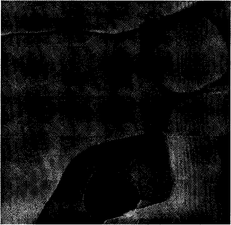
[0029] Example 1 A three-component lipid (DOPC / ESM / Chol) solid surface supported lipid film was prepared by spin coating.

[0030] The chloroform solutions of dioleoylphosphatidylcholine (DOPC), egg sphingolipid (ESM) and cholesterol (Chol) three kinds of phospholipids were mixed in a molar ratio of 2:2:1 to obtain lipid stock solution, wherein the concentration of lipid stock solution was 1.0mg (lipid) / mL (chloroform) solution. Use a spin coater to coat the mica surface with a stock solution, observe it under AFM at room temperature, and get image 3 . The parameters of the spin coater are: rotation speed 1000 rpm, spin coating time 20 s, and before AFM scanning, the sample is kept in a vacuum oven at 45°C. From image 3 It can be seen from the figure that the surface of the support film obtained by the spin coating method is not smooth, and the inside of the film is divided into several layers, which is not conducive to the observation and analysis of the morphology and i...

Embodiment 2

[0031] Example 2 The solid surface of the three-component lipid (DOPC / ESM / Chol) obtained by soaking in water and spin-coating supported the lipid film.

[0032]Soak the lipid film obtained in Example 1 above in Milli-Q water for 12 hours. After rinsing with Milli-Q water, it was placed in the AFM liquid pool and scanned at room temperature Figure 4 . It can be seen that even after the dry film is fully swollen in water, a flat surface cannot be obtained.

Embodiment 3

[0033] Example 3 A three-component lipid (DOPC / ESM / Chol) solid surface supported lipid membrane was prepared by common vesicle fusion method.

[0034] Take 2 mL of the stock solution in Example 1 and prepare it into a 1.0 mg (lipid) / mL (Milli-Q water) vesicle suspension solution. The average size of the vesicles is less than 200nm. Take 1mL of the vesicle suspension and put it on the surface of freshly peeled mica, keep it in an oven at 30°C for 4 hours, then carefully wash off the remaining solution with Milli-Q water, and rinse it 10 times repeatedly, trying to keep the surface in the water phase. The support membrane obtained after washing was placed in the AFM liquid pool for imaging, and the obtained Figure 5 . It can be seen that, compared with the supporting membranes obtained in Examples 1 and 2, the membranes prepared by the common vesicle fusion method are flat and have distinct phase boundaries. However, this method takes a long time for fusion, and at the same...


PUM


Abstract

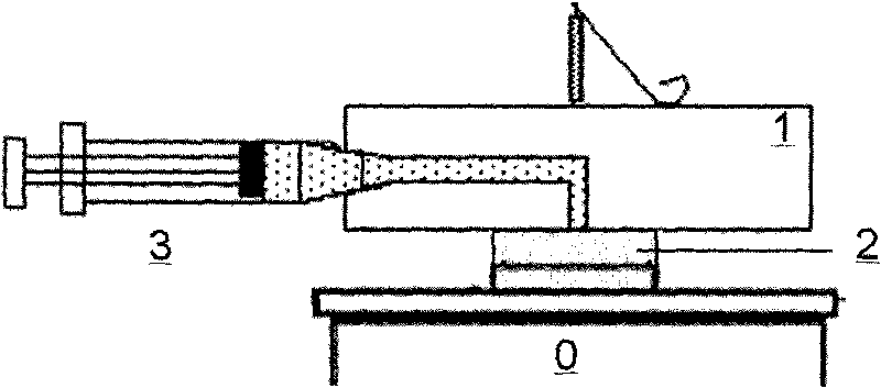
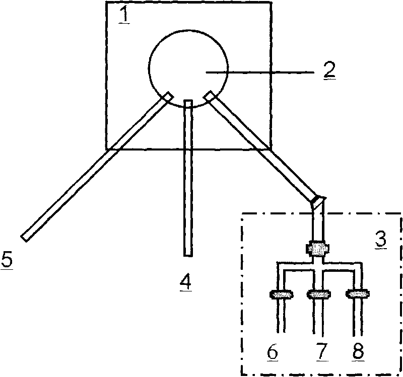
The invention belongs to the technical field of membrane preparation and particularly relates to a method and a device for preparing a lipid bilayer membrane in situ in a liquid phase. Different from the common vesicle fusion method, the in-situ high-temperature vesicle fusion method adopted by the invention is characterized in that: 1, a sample introduction device is designed by combining an atomic force microscope (AFM) liquid tank needle holder to prepare a bilayer membrane in situ on a sample table; and 2, the fusion process is implemented at the temperature higher than a room temperature, so the process is accelerated. The formed bilayer membrane can be used for the observation of and study on an atomic force microscope (AFM). The method not only enables the forming process and surface topography of the membrane to be monitored by the atomic force microscope in real time, but also can change the liquid-phase environment of the membrane in situ by controlling the sample introduction device so as to control chemical and physical adsorption, chemical reactions, crystal growth and the like on the surface of the membrane. The method also can be used for preparing and modifying other amphiphilic membranes.

Description

technical field [0001] The invention belongs to the technical field of membrane preparation, and in particular relates to a preparation method and device of a lipid bilayer membrane supported by a solid surface. Background technique [0002] The planar lipid bilayer (Supported Lipid Bilayer, SLB) supported by a solid surface is one of the main forms of model biological membranes. [1,2] By controlling the composition ratio of the lipid bilayer and the properties of the medium around the membrane and other influencing factors, people can use atomic force microscopy (AFM) to qualitatively and quantitatively study the response of biological membranes to external environmental changes and the internal mechanism of specific physiological processes. [2,3] The commonly used method of preparing planar support film is spin coating method. [3] , LB membrane transfer method [3,4] and vesicle fusion [1,3] Three categories. The spin coating method is to directly spin coat the organic ...

Claims


Application Information

Patent Timeline
Patent Type & Authority Applications(China)
IPC IPC(8): C04B41/47C03C17/28
Inventor 鲍稔李莉邱枫杨玉良
Owner FUDAN UNIV
Learn More