Monoclonal antibody for anti-human CEA, a composition containing same and application thereof
A technology of antibody and antibody light chain, applied in the field of molecular biology and biology
- Summary
- Abstract
- Description
- Claims
- Application Information
AI Technical Summary
Problems solved by technology
Method used
Image
Examples
Embodiment 1
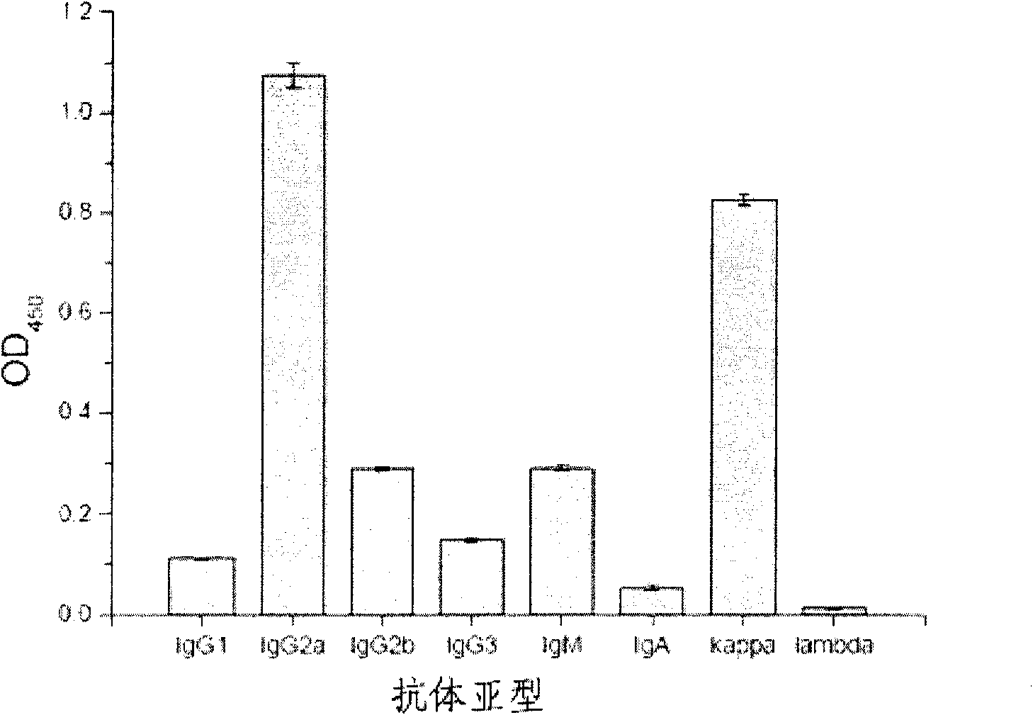
[0065] Example 1: Preparation and identification of monoclonal antibody CC4.
[0066] Application of hybridoma technology (Kohler and Milstein 1975; Yeh, Hellstrom et al.1979; Yeh, Hellstrom et al.1982) produced and screened to obtain antibody CC4. The brief description is as follows: BALB / c mice (Beijing Experimental Animal Center) were immunized with living human colorectal cancer cell LS 174T cells (ATCC) as antigen, and the mice were killed after 4 booster immunizations, and the spleen was taken and removed. Cells were suspended in RPMI medium. Splenocytes were fused with SP2 / 0-Ag14 murine myeloma cells (ATCC) in the presence of polyethylene glycol (PEG), and hybridomas were selected using HAT-selective medium. See reference (Kohler and Milstein 1975) for specific methods.
[0067] Using fixed LS 174T cells cultured in a 96-well cell culture plate as an antigen, the culture supernatants of hybridoma cells were screened by ELISA. The brief introduction of the cell ELISA ...
Embodiment 2
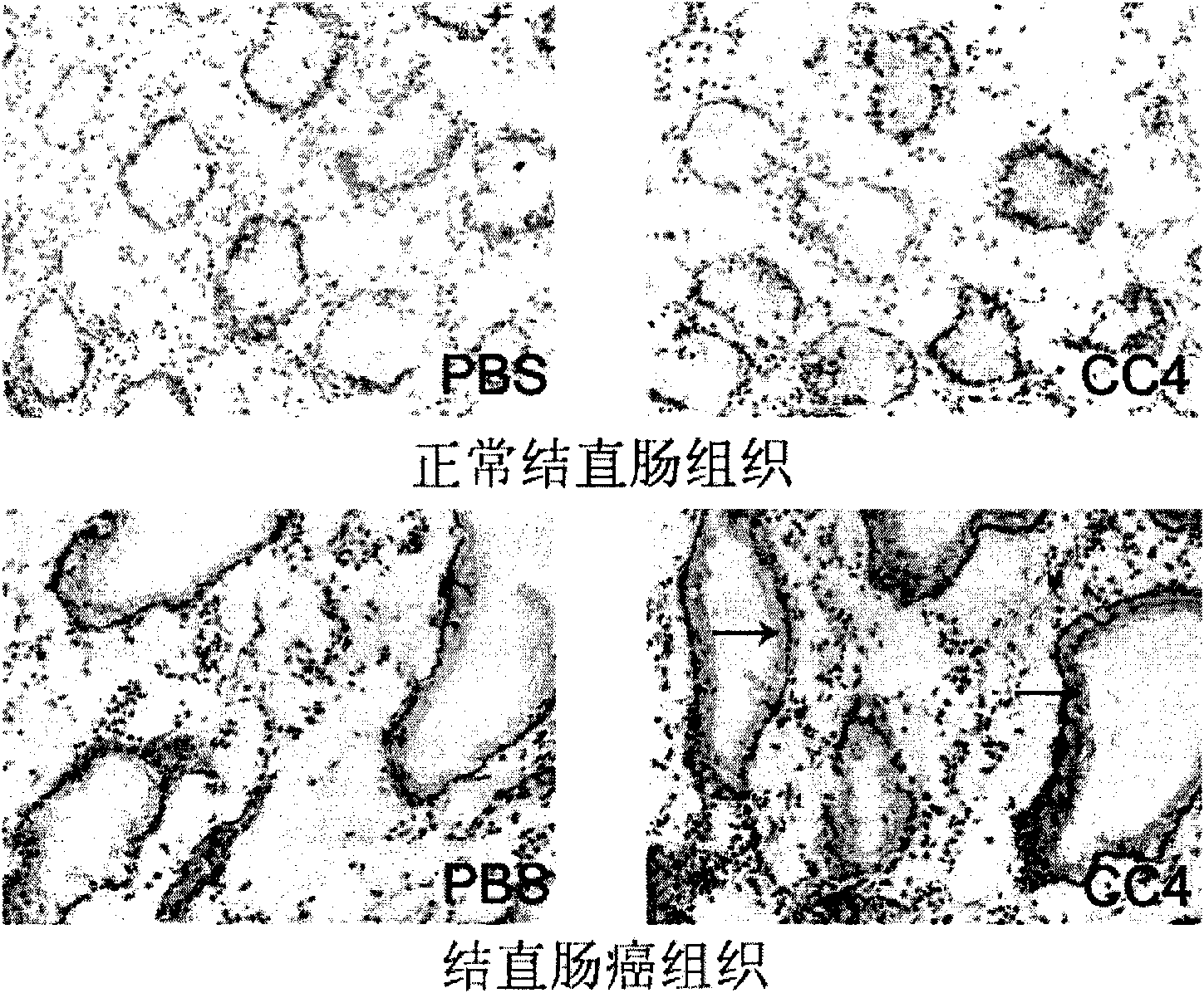
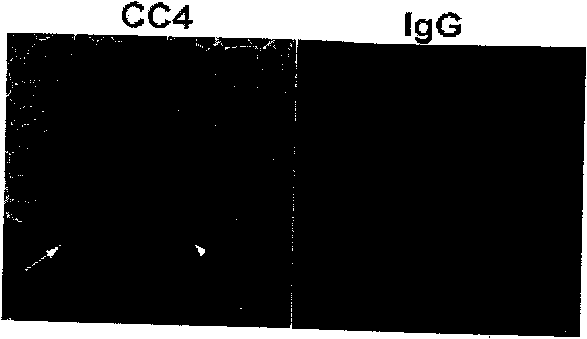
[0077] Example 2: Study on the specificity of monoclonal antibody CC4 to tumor tissue and tumor cells.
[0078] In order to systematically study the binding specificity of the monoclonal antibody CC4 of the present invention to human normal and tumor tissues, 29 cases of human normal tissues and 24 cases of frozen sections of human tumor tissues were screened by immunohistochemical method.
[0079] The specific experimental method is as follows: human normal tissue or tumor tissue was embedded with OCT (frozen tissue embedding fluid, provided by Sakura), and frozen section was performed. Sections were fixed in pre-cooled acetone for 5 min, then in 0.3% HO 2 o 2 Incubate in dark methanol at room temperature for 30 minutes to remove the interference of endogenous peroxidase. After washing with PBS for three times, the sections were blocked with 5% horse serum (Beijing Zhongshan Jinqiao, horse serum diluted in PBS), and then in the primary antibody dilution solution (1:2000 blo...
Embodiment 3
[0097] Example 3: Antigen identification of monoclonal antibody CC4.
[0098] Using western blotting, it was found that CC4 recognized a 130kD protein present in LS 174T cells and colorectal cancer tissues. The specific operation is as follows: collect the human-derived colorectal cancer cell LS 174T (ATCC) that strongly binds to CC4, wash the cells twice with pre-cooled PBS, centrifuge at 800 rpm at 4°C for 5 minutes, and use lysate (Tris-HCl 50mM pH 8.0, NaCl 150mM, EDTA 1mM, NP-40 1%, Glycerol 10%, PMSF 100μg / ml) to lyse the cells, centrifuge at 12000g at 4°C for 15 minutes, collect the supernatant, add DTT (final concentration 100mM) (dithiothreose Alcohol) and DTT-free loading buffer (5x loading buffer: 0.313M Tris-HCl, pH6.8, 10% SDS, 0.05% bromophenol blue, 50% glycerol), cook at 100°C. Whole-cell proteins were separated by 10% SDS-PAGE, followed by semi-dry electrotransfer to nitrocellulose membranes. After blocking with 5% skimmed milk, CC4 ascites was used as the p...
PUM
 Abstract
Description
Claims
Application Information
 - R&D
- Intellectual Property
- Life Sciences
- Materials
- Tech Scout
- Unparalleled Data Quality
- Higher Quality Content
- 60% Fewer Hallucinations
Browse by: Latest US Patents, China's latest patents, Technical Efficacy Thesaurus, Application Domain, Technology Topic, Popular Technical Reports.
© 2025 PatSnap. All rights reserved.Legal|Privacy policy|Modern Slavery Act Transparency Statement|Sitemap|About US| Contact US: help@patsnap.com


