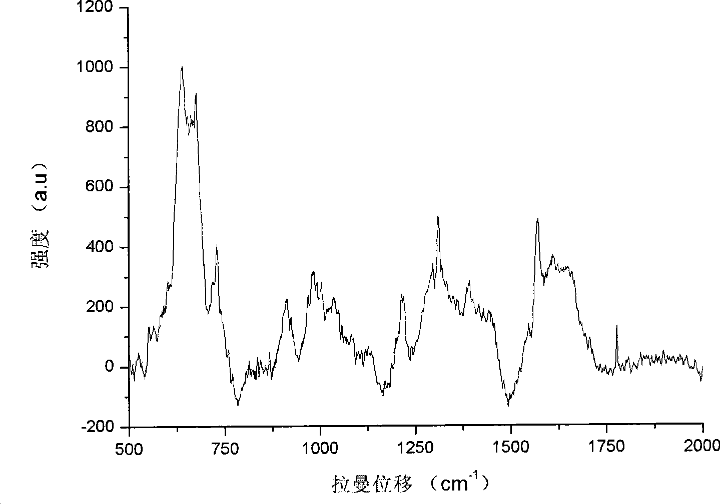
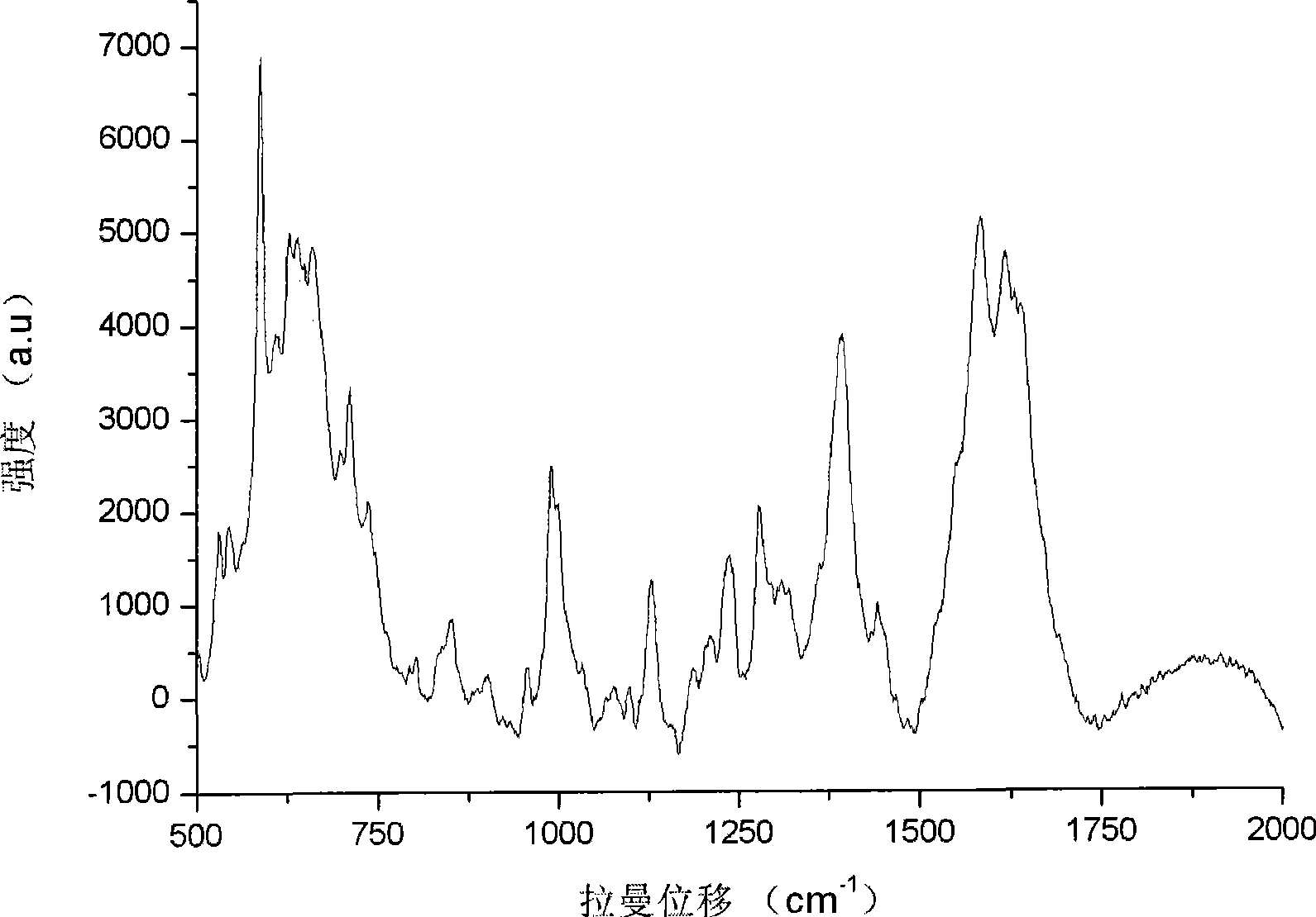
Method for detecting animal active unicellular sample by surface reinforced Raman spectrum
A surface-enhanced Raman and spectroscopic detection technology, applied in the field of biomedicine, can solve the problems of micropores in the cell membrane, decrease, and the cell membrane hinders the permeability of particles, and achieves the effect of high transfer efficiency and a simple method.
- Summary
- Abstract
- Description
- Claims
- Application Information
AI Technical Summary
Problems solved by technology
Method used
Image
Examples
Embodiment 1
[0021] Dissolve 9mg of silver nitrate in 50ml of deionized water, heat to 100°C to make it boil, then add 1ml of 3% trisodium citrate dropwise, stir rapidly, and keep boiling for 2 hours after the dropwise addition. After the silver sol was naturally cooled, the layers were centrifuged in a centrifuge, the supernatant was discarded, and the concentrated silver sol in the lower layer was sealed and stored at room temperature away from light for later use.
[0022] The test sample epithelial tumor cells (A431) to be tested are first prepared with commercially available RPMI 1640 cell culture medium as a single-cell suspension of epithelial tumor cells, and then the cell content is calculated by a hemocytometer, and the cell density is controlled to be 10 5 pieces / ml.
[0023] Take 80 μl and 320 μl of single-cell suspensions of concentrated silver sol and epithelial tumor cells (A431), mix them at room temperature, and transfer them to an electric shock cup sample pool. The elect...
Embodiment 2
[0025] Dissolve 12mg of silver nitrate in 50ml of deionized water, heat to 100°C to make it boil, then add 1ml of 1% trisodium citrate dropwise, stir rapidly, and keep boiling for 4 hours after the dropwise addition. After the silver sol was naturally cooled, the layers were centrifuged in a centrifuge, the supernatant was discarded, and the concentrated silver sol in the lower layer was sealed and stored at room temperature away from light for later use.
[0026] The test sample leukemia tumor cells (HL60) to be tested are first prepared with commercially available RPMI 1640 cell culture medium as a single-cell suspension of epithelial tumor cells, and then the cell content is calculated by a hemocytometer, and the cell density is controlled to be 10 6 pieces / ml.
[0027] Take concentrated silver sol and 120 μl and 480 μl of single-cell suspension of leukemia tumor cells (HL60) respectively, mix them at room temperature, and transfer them into an electric shock cup sample poo...
Embodiment 3
[0029] Dissolve 10mg of silver nitrate in 50ml of deionized water, heat to 100°C to make it boil, then add 1ml of 2% trisodium citrate dropwise, stir rapidly, and keep boiling for 6 hours after the dropwise addition. After the silver sol was naturally cooled, the layers were centrifuged in a centrifuge, the supernatant was discarded, and the concentrated silver sol in the lower layer was sealed and stored at room temperature away from light for later use.
[0030] The test sample nasopharyngeal carcinoma tumor cells (C66) to be tested are first made into a single-cell suspension of epithelial tumor cells with commercially available RPMI 1640 cell culture medium, and then the content of the cells is calculated by a hemocytometer, and the control cell density is 10 5 pieces / ml.
[0031] Take concentrated silver sol and 160 μl and 640 μl of single cell suspension of nasopharyngeal carcinoma tumor cells (C66) and mix them at room temperature, then transfer them to an electric sho...
PUM
 Abstract
Description
Claims
Application Information
 - R&D Engineer
- R&D Manager
- IP Professional
- Industry Leading Data Capabilities
- Powerful AI technology
- Patent DNA Extraction
Browse by: Latest US Patents, China's latest patents, Technical Efficacy Thesaurus, Application Domain, Technology Topic, Popular Technical Reports.
© 2024 PatSnap. All rights reserved.Legal|Privacy policy|Modern Slavery Act Transparency Statement|Sitemap|About US| Contact US: help@patsnap.com


