
Device for superposed mri and pet imaging

A device, a technology of magnetic resonance tomography, applied in the direction of instruments used for radiological diagnosis, measurement using nuclear magnetic resonance imaging system, application, etc., can solve the problems of large location requirements, high cost, no possibility of shielding PET detectors, etc.

Active Publication Date: 2008-09-03
SIEMENS HEATHCARE GMBH
View PDF0 Cites 13 Cited by
  • Summary
  • Abstract
  • Description
  • Claims
  • Application Information

AI Technical Summary

Problems solved by technology

[0012] Finally, based on the radial positional conditions, there is no possibility at all to shield (for example, through the septum) the ring-shaped PET detector from gamma rays outside the ring-shaped PET detector
[0013] A solution such as highly integrated or linked HF systems and PET detectors, resulting in higher costs for the mechanics and higher costs in the event of failure
Another workaround is to basically unintegrate, as in Image 6 as shown in the reference example, but the resulting relatively large location requirements

Method used


Image

  • Device for superposed mri and pet imaging
  • Device for superposed mri and pet imaging
  • Device for superposed mri and pet imaging

Examples


Embodiment Construction

[0034] Embodiments of the present invention will be described below with reference to the accompanying drawings.

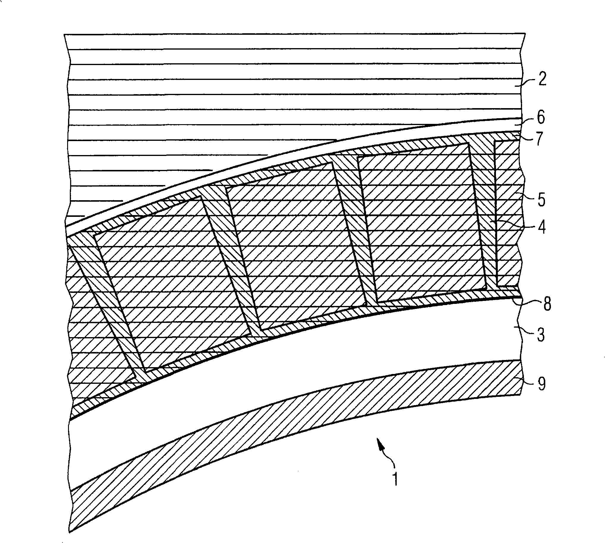
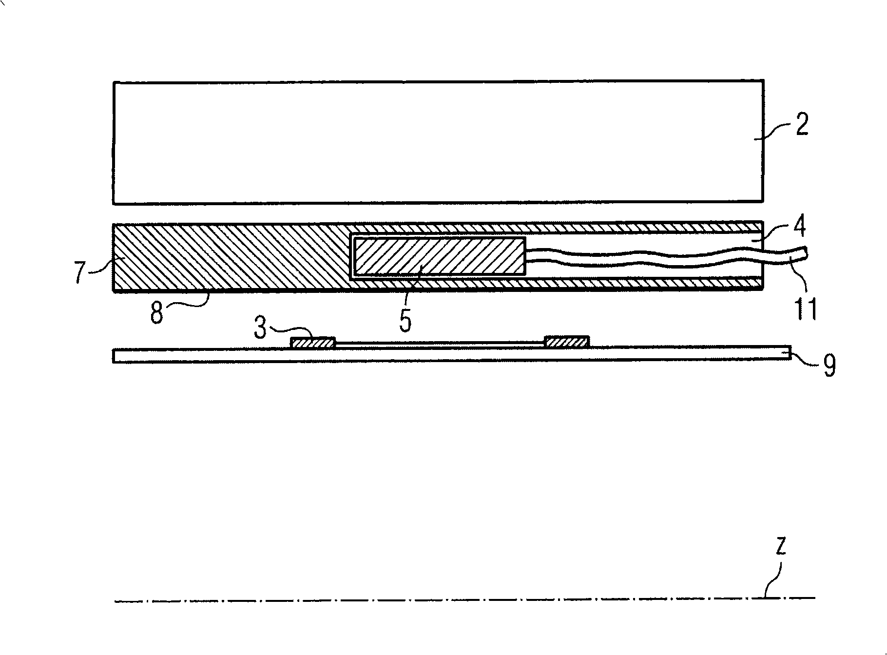
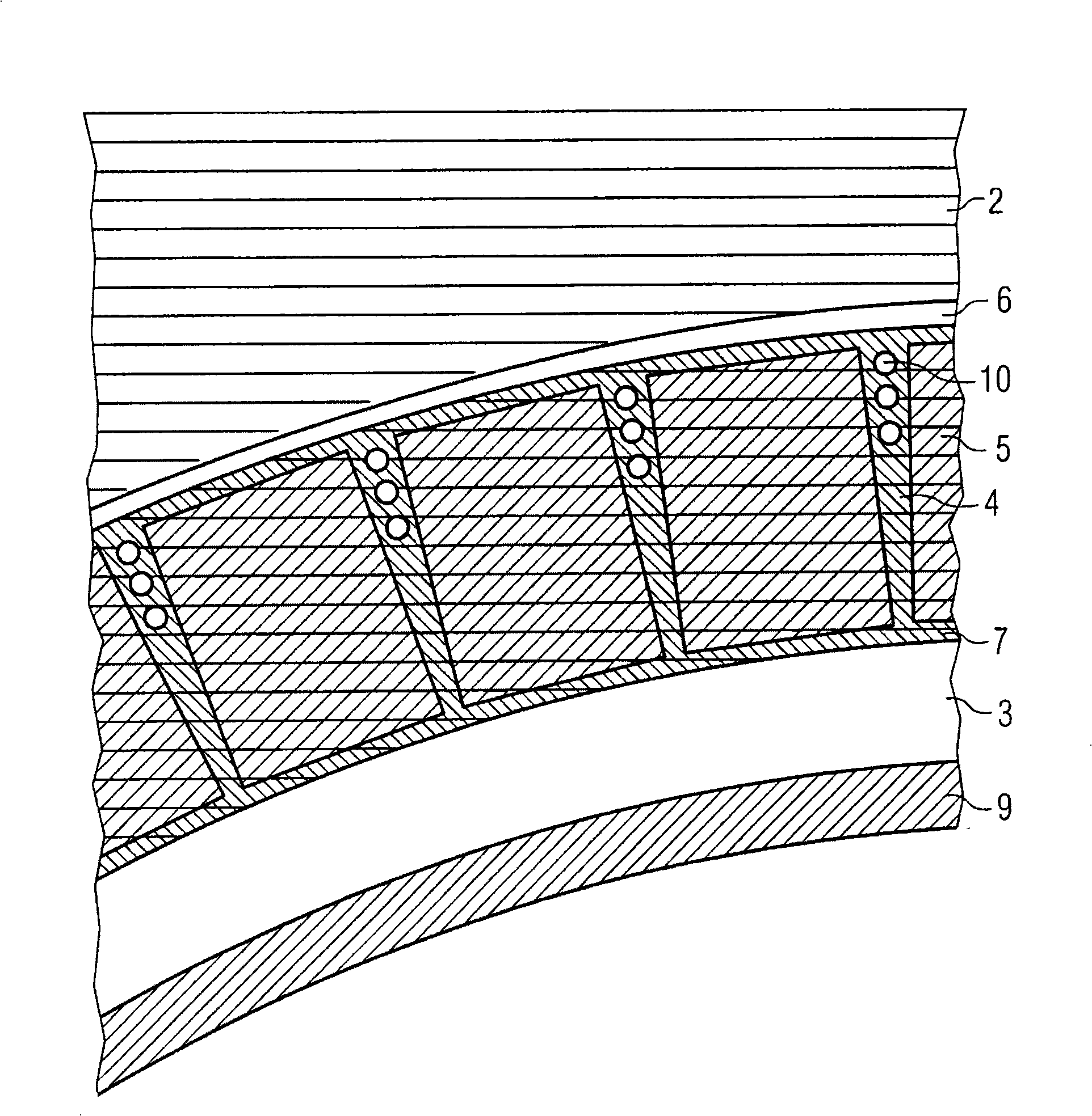
[0035] figure 1 shows an apparatus for superimposing a magnetic resonance tomography image and a positron emission tomography image according to a first embodiment of the present invention, and figure 2 Device according to the invention for superimposing magnetic resonance tomography images and positron emission tomography images figure 1 Longitudinal sectional view of the part shown.

[0036] According to the invention, the device 1 for superimposing magnetic resonance tomography images and positron emission tomography images has a magnetic resonance tomography magnet (not shown), which is defined as Figure 5 The longitudinal axis z indicated in. Magnetic resonance tomography magnets form a basic field system, which provides a strong static magnetic field.

[0037] Furthermore, the device 1 has a magnetic resonance tomography gradient coil 2 arranged radia...


PUM


Abstract

A device is disclosed for superposed magnetic resonance imaging and positron emission tomography imaging. The device includes a magnetic resonance imaging magnet which defines a longitudinal axis (z); a magnetic resonance imaging gradient coil (2) arranged radially within the magnetic resonance imaging magnet; a magnetic resonance imaging HF coil (3) arranged radially within the magnetic resonance imaging gradient coil (2); and a multiplicity of positron emission tomography detection units arranged in pairs opposite one another about the longitudinal axis (z). The many positron emission tomography detection units (5) are arranged radially within the magnetic resonance imaging gradient coil (2) and can be inserted into the device and can be removed from the device along the longitudinal axis (z). A carrier tube (7) is preferably provided having a multiplicity of pockets (4), which in each case extend along the longitudinal axis, for accommodating at least one positron emission tomography detection unit (5).

Description

technical field [0001] The invention relates to a device for superimposing MRI images and PET images. Background technique [0002] Magnetic resonance tomography (MRI apparatus) is known to have three main functional parts, which are represented in Figure 5 In the center: the fundamental field system 21 , the gradient system 22 and the high frequency system 23 (also called HF system or body resonator). The basic field system 21 is usually a magnet and provides a strong static magnetic field. The gradient system 22 provides a tunable magnetic field in the low frequency range down to about 1 kHz, which has a linear ramp up or down in one or more directions. The HF system 23 provides an oscillating magnetic field for deflecting nuclear spins in the high frequency range substantially at the nuclear spin resonance frequency (typically 42.45 MHz) given by the static magnetic field, and it can also be used to receive Yu's nuclear spin signal. [0003] In a common magnetic reson...

Claims


Application Information

Patent Timeline
IPC IPC(8): A61B5/055A61B6/03G01R33/48
CPCG01R33/3856A61B6/4417A61B6/037G01R33/481G01T1/1603
Inventor 于尔根·尼斯特勒沃尔夫冈·伦兹洛瑟·舍恩斯蒂芬·斯托克
Owner SIEMENS HEATHCARE GMBH
Learn More