
Method and apparatus for virus and vaccine production

a technology for vaccines and methods, applied in the field of methods and equipment for virus and vaccine production, can solve the problems of high impurity levels, low yield and high impurity levels, and is difficult to produce efficiently with traditional large-scale manufacturing techniques, etc., to achieve cost-effectiveness, eliminate the need for cleaning and reuse, and facilitate direct applicability to therapies and production

Active Publication Date: 2017-08-15
BIOVEST INT
View PDF35 Cites 15 Cited by
  • Summary
  • Abstract
  • Description
  • Claims
  • Application Information

AI Technical Summary

Benefits of technology

[0019]In some embodiments, the bioreactor is a hollow fiber bioreactor in which the first compartment is an intracapillary (IC) space, the second compartment is an extracapillary (EC) space, the membrane is a hollow fiber membrane, the virus-infected cells are cultured in the IC space or the EC space, and the cells are cultured under conditions that allow for regulating (e.g., reducing) the concentration of molecules inhibitory to virus or VLP yield within the IC space or the EC space, and allow for production of the virus or VLP at a yield greater than that achieved in the absence of said regulation. Virus-infected cells may be cultured within the EC space or the IC space of the bioreactor. In some embodiments, virus-infected cells are cultured in the EC space and the concentration of inhibitory molecules within the EC space is regulated by increasing the flow rate of culture media on the EC side (EC circuit) of the bioreactor and controlling how fast culture media bearing the inhibitory molecules is replaced with fresh culture media on the EC side of the bioreactor.
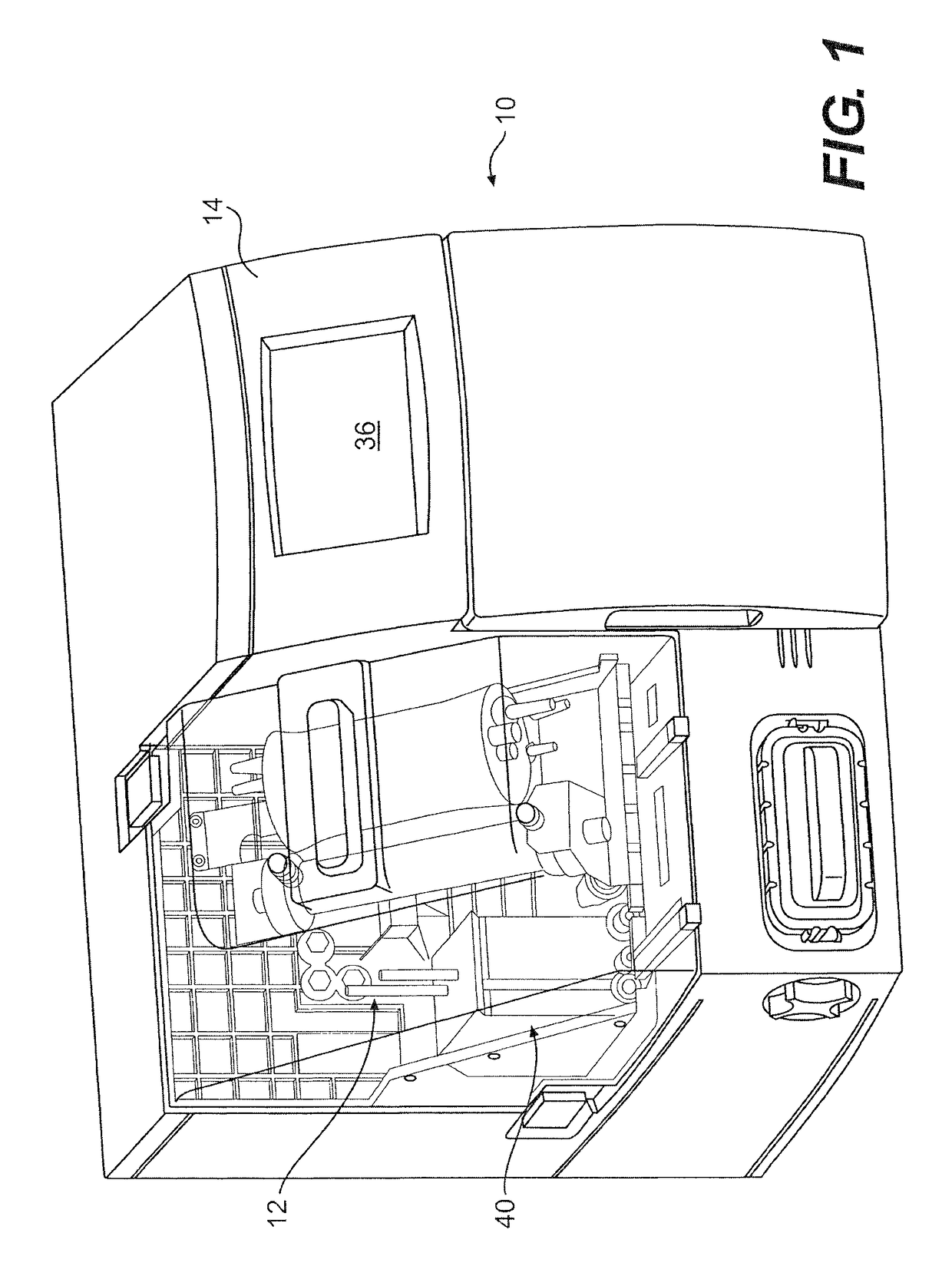
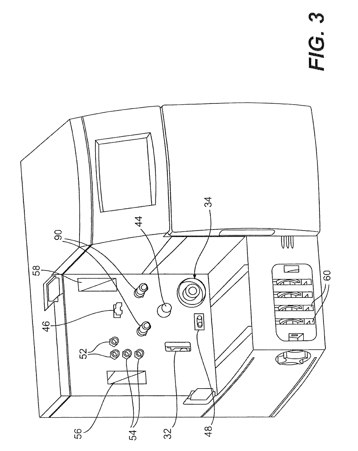
[0038]Any host cells useful for production of the virus of interest can be used in the methods, apparatus, systems, and bioreactors of the invention. Host cells may be human or non-human mammalian cells, insect cells, avian cells, or plant cells. For example, Madin-Darby canine kidney (MDCK) cells and African green monkey kidney (Vero) cells are useful for the production of influenza virus. Other continuous cell lines suitable as host cells for production of human vaccines include human fetal retina (Per.C6) and duck embryonic retina (AGE1.CR) cells. The host cells may be transformed or non-transformed cell lines, primary cells including somatic cells such as lymphocytes or other immune cells, chondrocytes, myocytes or myoblasts, epithelial cells and patient specific cells, primary or otherwise. Included also are cells or cell lines that have been genetically modified, such as both adult and embryonic stem cells. The automated cell culture apparatus allows for production and harvest of cells or cell-secreted protein in a manner that minimizes the need for operator intervention and minimizes the need for segregated clean rooms for the growth and manipulation of the cells. Further, the apparatus provides a culture environment that is completely self-contained and disposable. This eliminates the need for individual clean rooms typically required in a regulated, multi-use facility. Control of fluid dynamics within the bioreactor allows for growth conditions to be adjusted, e.g., changing growth factor concentrations, to facilitate application of unique culture protocols or expansion of unique cells or cell lines. As a result, there is less variation and less labor required for consistent, reproducible production of cells and viral products for applications to expansion of cells.

Problems solved by technology

The greatest challenge is expected to come in the area of virus-based therapies such as vaccines and viral vectors for gene therapy.
Traditional methods for virus purification, by their nature, are prone to technician error, inefficiency or inconsistency leading to low yield and high levels of impurities.
Patient-specific therapies require relatively small quantities of the therapeutic agent and such small quantities are impossible to produce efficiently with traditional large scale manufacturing techniques.
For example, these methods can be labor intensive, time consuming, and are highly inefficient.
The manual steps involved in these methods also include a high risk of contamination.
Moreover, conventional approaches and tools for viruses or virus-based products typically involve numerous manual manipulations that are subject to variations even when conducted by skilled technicians.
When used at the scale needed to manufacture hundreds or thousands of different cells, cell lines and patient-specific cell based therapies, the variability, error or contamination rate may become unacceptable for commercial processes.
These methods are very labor intensive, have inconsistent performance, are subject to mistakes, and difficult for large-scale production.
This method is also very labor intensive, difficult for large scale production, and objectionable because of the potential for egg-based allergic reactions and variable productivity.
This method is costly in terms of facility support in order to accommodate a large number of unique cells and produces product at low concentration.
This can bring facilities costs down and increases product concentration.
The systems currently available are general purpose in nature and require considerable time from trained operators to setup, load, flush, inoculate, run, harvest, and unload.
Each step typically requires manual documentation, which is labor intensive and subject to errors.
Consequently, compactness of the design and the amount of ancillary support resources required will become an important facilities issue.
Moreover, the systems currently available are general purpose in nature and require considerable time from trained operators to setup, load, flush, inoculate, run, harvest and unload.
This is labor intensive and subject to errors.
This is a time intensive activity.
While well-understood, robust and convenient, classical batch-style two-dimensional (2-D) culture on non-porous supports or three-dimensional (3-D) suspension culture in other devices do not replicate in vivo conditions and may affect protein modifications.

Method used


Image

  • Method and apparatus for virus and vaccine production
  • Method and apparatus for virus and vaccine production
  • Method and apparatus for virus and vaccine production

Examples


embodiment 1

[0093]A method for the production of virus and virus-like particles (VLP), comprising culturing virus-infected cells in a cell culture bioreactor comprising a first compartment, a second compartment, and a membrane separating the first and second compartments, wherein the cells are cultured in the first compartment under conditions that allow for regulating the concentration of a molecule inhibitory to virus or VLP yield from the cells within the first compartment, and allow for production of the virus or VLP at a yield greater than that achieved in the absence of said regulation.

embodiment 2

[0094]The method of embodiment 1, wherein the bioreactor is a hollow fiber bioreactor, the first compartment is an extracapillary (EC) space, the second compartment is an intracapillary (IC) space, and the membrane comprises a hollow fiber membrane.

embodiment 3

[0095]The method of embodiment 1, wherein the bioreactor is a hollow fiber bioreactor, the first compartment is intracapillary (IC) space, the second compartment is an extracapillary (EC) space, and the membrane comprises a hollow fiber membrane.


PUM

PropertyMeasurementUnit
temperatureaaaaaaaaaa
diameteraaaaaaaaaa
pore sizeaaaaaaaaaa

Abstract

The invention concerns a bioreactor for production of virus and virus-like particles (VLPs), methods for production of virus and VLPs, methods for regulating the concentration of molecules inhibitory to viral and VLP yield in a cell culture chamber of a bioreactor, such as the extracapillary space of a hollow fiber bioreactor.

Description

CROSS-REFERENCE TO RELATED APPLICATIONS[0001]This application is a continuation of U.S. application Ser. No. 14 / 122,309, filed Nov. 26, 2013, which is the National Stage of International Application No. PCT / US2012 / 041942, filed Jun. 11, 2012, which claims the benefit of U.S. Provisional Application Ser. No. 61 / 495,882, filed Jun. 10, 2011, U.S. Provisional Application Ser. No. 61 / 496,432, filed Jun. 13, 2011, and U.S. Provisional Application Ser. No. 61 / 496,898, filed Jun. 14, 2011, the disclosure of each of which is incorporated herein by reference in its entirety, including all figures, tables and drawings.BACKGROUND OF THE INVENTION[0002]The anticipated growth of cell culture-based virus production will require new paradigms for rapid, high-throughput, harvest, purification, concentration and formulation of a variety of human or non-human viruses. The greatest challenge is expected to come in the area of virus-based therapies such as vaccines and viral vectors for gene therapy. I...

Claims


Application Information

Patent Timeline
Patent Type & Authority Patents(United States)
IPC IPC(8): C12N7/00C12M1/12C12M3/00C12M1/00C12M1/36C12M1/34
CPCC12M23/28C12M23/34C12M23/44C12M25/10C12M29/04C12M41/12C12M41/32C12M41/48C12M47/12C12N7/00C12N2760/16051
Inventor HIRSCHEL, MARKGANGEMI, JOSEPH DAVID
Owner BIOVEST INT
Learn More